

doi: 10.1016/j.jneuroim.2009.06.016.
Epub 2009 Jul 28.
Mapping of the full length and the truncated interleukin-18 receptor alpha in the mouse brain
Affiliations
- PMID: 19640592
- PMCID: PMC2745497
- DOI: 10.1016/j.jneuroim.2009.06.016
Item in Clipboard
Mapping of the full length and the truncated interleukin-18 receptor alpha in the mouse brain
J Neuroimmunol.
.
Abstract
The cytokine IL-18 acts on the CNS both in physiological and pathological conditions. Its action occurs through the heterodimeric receptor IL-18Ralpha\beta. To better understand IL-18 central effects, we investigated in the mouse brain the distribution of two IL-18Ralpha transcripts, a full length and an isoform lacking the intracellular domain hypothesized to be a decoy receptor. Both isoforms were expressed in neurons throughout the brain primarily with overlapping distribution but also with some unique pattern. These data suggest that IL-18 may modulate neuronal functions and that its action may be regulated through expression of a decoy receptor.
Figures
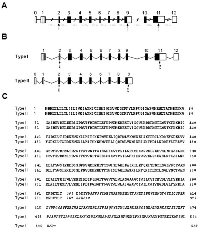
(A) Arrangement of exons and introns, drawn to scale, of mouse IL-18Rα gene structure as determined by analyzing genomic and mRNAs sequences. Exons are shown as boxes and introns are shown as lines. (B) Schematic representation of IL-18Rα transcripts in relation to the gene. Unranslated regions (UTR) are shown as empty boxes and coding sequences (CDS) are shown as black boxes. The putative translation initiation codon (ATG) and the termination codon (STOP) are also indicated. (C) Alignment of the amino acid sequences of the canonical and short isoforms with the predicted transmembrane regions underlined while specific type I or type II amino acid sequences are indicated in italic font.
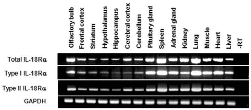
GAPDH mRNA was measured as an internal control. The lengths of the expected PCR products were: 342 bp, 215 bp, 240 bp and 89 bp for total, type I, type II IL-18Rα and GAPDH respectively. Total RNA was extracted from brain areas and tissues of 3 mice and pooled before RT reaction.
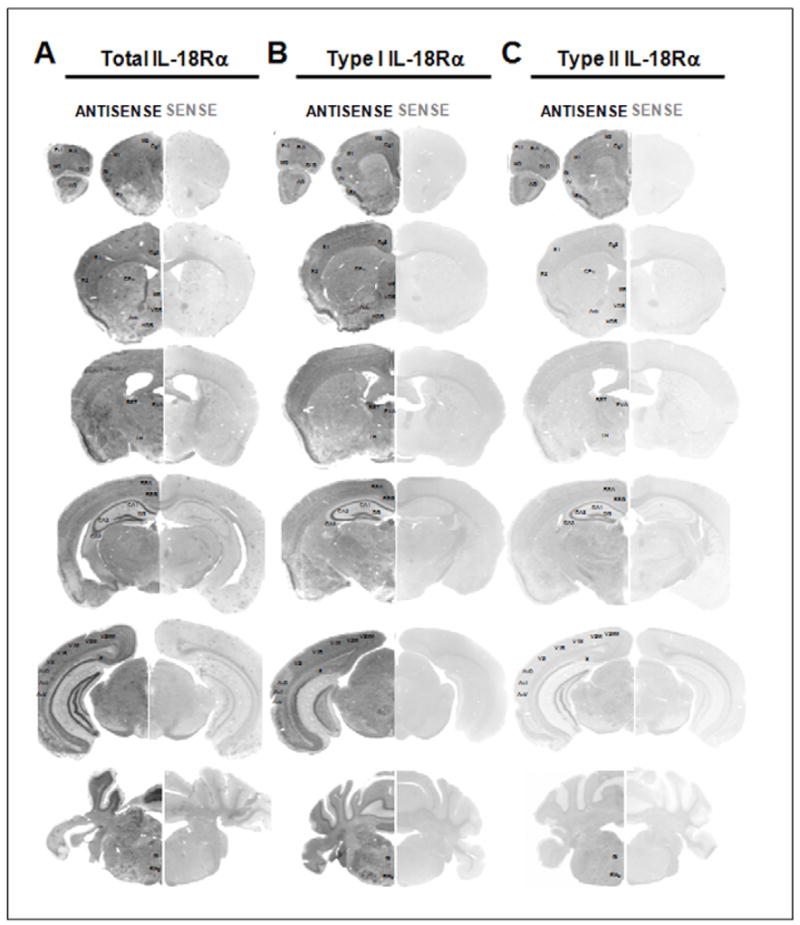
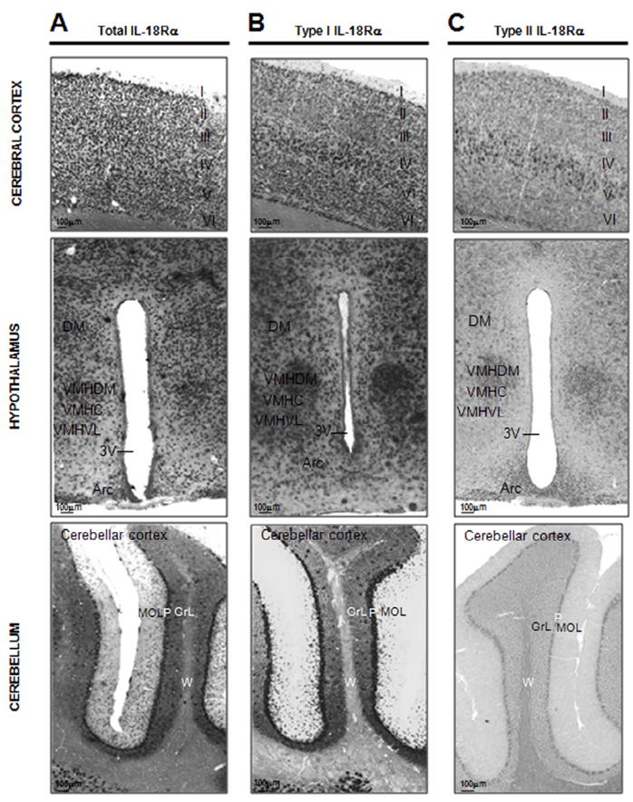
Expression of the mRNAs of interest demonstrated by using in situ hybridization with a DIG-labeled antisense cRNA specific probes (ANTISENSE lanes) and using a sense cRNA probes (SENSE lanes) as a negative control. Section hybridized with the sense riboprobes showing the absence of hybridization signal (SENSE lanes). Acb, nucleus accumbens; AI, agranular insular cortex; AO, anterior olfactory nuclei; Au1, primary auditory cortex; AuD, auditory cortex, dorsal part; AuV, auditory cortex, ventral part; BST, bed nucleus of the stria terminalis; CA, Ammon’s horn; CA1, CA1 field of the hippocampus; CA2, CA2 field of the hippocampus; CA3, CA3 field of the hippocampus; Cg1, cingulate cortex area 1; Cg2, cingulate cortex area 2; CPu, caudate-putamen; DG, dentate gyrus; DLO, dorsolateral orbital cortex; FrA, frontal association cortex; Gi, gigantocellular reticular nucleus; GI, granular insular cortex; HDB, nucleus of the horizontal limb of the diagonal band; LH, lateral hypothalamic area; M1, primary motor cortex; M2, secondary motor cortex; MO, medial orbital cortex; MS, medial septal nucleus; Pir, piriform cortex; PrL, prelimbic cortex; PVA, paraventricular thalamic nucleus anterior part; RMg, raphe magnus nucleus. RSA, retrosplenial agranular cortex; RSG, retrosplenial granular cortex; S, subiculum; S1, primary somatosensory cortex; S2, secondary somatosensory cortex; V1B, primary visual cortex, binocular region; V1M, primary visual cortex, monocular region; V2L, secondary visual cortex, lateral part; V2ML, visual cortex 2, mediolateral part; V2MM, visual cortex 2, mediodedial part; VDB, nucleus of the vertical limb of the diagonal band.
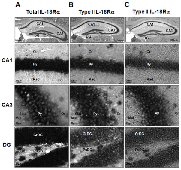
Shown are representative coronal brain sections at the level of the dorsal hippocampus exhibiting in situ hybridization with the indicated digoxigenin-labeled riboprobes. CA1: CA1 field of the hippocampus, CA3: CA3 field of the hippocampus, DG: dentate gyrus, Or: oriens layer of the hippocampus, Py: pyramidal cell layer of the hippocampus, Rad: stratum radiatum of the hippocampus, SLu: stratum lucidem of the hippocampus, Mol: molecular layer of the dentate gyrus, GrDG: granular layer of the dentate gyrus, PoDG: polymorph layer of the dentate gyrus.
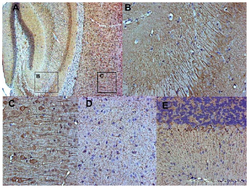
Animals were perfused, the brains were harvestedand embedded in paraffin. The IL-18Rα receptor was detected using Mab 1216 (R&D) on 5 μm sections. Negative controls were performed by omitting the primary antibody, confirming the specificity of the antibody binding. (A), (B) and (C) Staining pattern using antibody sc-34178, showing predominantly neuronal cell bodies. (D) and E) Staining pattern using antibody Mab 1216, showing mostly dendrites. (A) Specific staining (brown color) on cortex and hippocampus (8x magnification). Rectangles indicate areas presented in detail in (B) and (C); (B) IL18Ra-positive cells (dark brown) in the dentate gyrus (DG) (32x); (C) Cortical detail, revealing a staining pattern associated to cell bodies and predominantly dendrites (32x). (D) Detailed view of neurons in the anterior hypothalamus (32x), and (E) cerebellum, showing positive cells (brown), associated exclusively to dendrites on Purkinje cells (20x).
References
-
- Andoh T, Kishi H, Motoki K, Nakanishi K, Kuraishi Y, Muraguchi A. Protective effect of IL-18 on kainate- and IL-1 beta-induced cerebellar ataxia in mice. J Immunol. 2008;180:2322–2328. - PubMed
-
- Andre R, Wheeler RD, Collins PD, Luheshi GN, Pickering-Brown S, Kimber I, Rothwell NJ, Pinteaux E. Identification of a truncated IL-18R beta mRNA: a putative regulator of IL-18 expressed in rat brain. J Neuroimmunol. 2003;145:40–45. - PubMed
-
- Bossu P, Ciaramella A, Moro ML, Bellincampi L, Bernardini S, Federici G, Trequattrini A, Macciardi F, Spoletini I, Di Iulio F, Caltagirone C, Spalletta G. Interleukin 18 gene polymorphisms predict risk and outcome of Alzheimer’s disease. J Neurol Neurosurg Psychiatry. 2007;78:807–811. - PMC - PubMed
-
- Bourke E, Cassetti A, Villa A, Fadlon E, Colotta F, Mantovani A. IL-1 beta scavenging by the type II IL-1 decoy receptor in human neutrophils. J Immunol. 2003;170:5999–6005. - PubMed
-
- Colotta F, Re F, Muzio M, Bertini R, Polentarutti N, Sironi M, Giri JG, Dower SK, Sims JE, Mantovani A. Interleukin-1 type II receptor: a decoy target for IL-1 that is regulated by IL-4. Science. 1993;261:472–475. - PubMed
Publication types
MeSH terms
Substances
Grants and funding
LinkOut - more resources
Full Text Sources
Miscellaneous
