. 2010 Mar 6;7(44):453-65.
doi: 10.1098/rsif.2009.0255. Epub 2009 Jul 29.

Effect of nanoparticulate bioactive glass particles on bioactivity and cytocompatibility of poly(3-hydroxybutyrate) composites

Affiliations

Effect of nanoparticulate bioactive glass particles on bioactivity and cytocompatibility of poly(3-hydroxybutyrate) composites

Superb K Misra et al. J R Soc Interface. .

Abstract

This work investigated the effect of adding nanoparticulate (29 nm) bioactive glass particles on the bioactivity, degradation and in vitro cytocompatibility of poly(3-hydroxybutyrate) (P(3HB)) composites/nano-sized bioactive glass (n-BG). Two different concentrations (10 and 20 wt %) of nanoscale bioactive glass particles of 45S5 Bioglass composition were used to prepare composite films. Several techniques (Raman spectroscopy, scanning electron microscopy, atomic force microscopy, energy dispersive X-ray) were used to monitor their surface and bioreactivity over a 45-day period of immersion in simulated body fluid (SBF). All results suggested the P(3HB)/n-BG composites to be highly bioactive, confirmed by the formation of hydroxyapatite on material surfaces upon immersion in SBF. The weight loss and water uptake were found to increase on increasing bioactive glass content. Cytocompatibility study (cell proliferation, cell attachment, alkaline phosphatase activity and osteocalcin production) using human MG-63 osteoblast-like cells in osteogenic and non-osteogenic medium showed that the composite substrates are suitable for cell attachment, proliferation and differentiation.


Figures

Figure 1.
Figure 1.
SEM micrographs of (a) P(3HB), (b) P(3HB)/10 wt % n-BG and (c) P(3HB)/20 wt % n-BG.
Figure 2.
Figure 2.
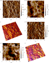
AFM images over a scan area of 10 × 10 µm2 showing the (a) amplitude channel for P(3HB) film, (b) height channel for P(3HB) film, (c) three-dimensional image of P(3HB) film showing the height and the phase channel superimposed, (d) amplitude channel for P(3HB)/20 wt % n-BG film, (e) height channel for P(3HB)/20 wt % n-BG film and (f) three-dimensional image of P(3HB)/20 wt % n-BG film showing the height and the phase channel superimposed.
Figure 3.
Figure 3.
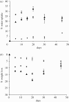
In vitro degradation study of P(3HB)/n-BG composites in SBF depicting (a) % water uptake and (b) % weight loss. Diamonds, P(3HB); squares, 10 wt % BG; triangles, 20 wt % BG.
Figure 4.
Figure 4.
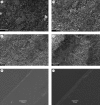
SEM micrographs of P(3HB)/20 wt % n-BG composite samples upon immersion in SBF showing the surface changes owing to the formation of HA layer after (a) 5 days (scale bar, 5 µm), (b) 15 days (scale bar, 10 µm), (c) 30 days (scale bar, 10 µm) and (d) 45 days (scale bar, 100 µm). Cross sections of P(3HB)/n-BG films showing the presence of continuous HA layer after 30 days in SBF for (e) P(3HB)/10 wt % n-BG (scale bar, 50 µm) and (f) P(3HB)/20 wt % n-BG (scale bar, 20 µm).
Figure 5.
Figure 5.
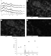
(a) XRD pattern of P(3HB)/n-BG composites highlighting the emergence of crystalline HA peak (denoted by vertical markers) after 5, 20 and 30 days of immersion in SBF. Results of EDX analysis for P(3HB)/20 wt % n-BG composites after 30 days in SBF showing elemental surface mapping of (b) Ca, (c) P, (d) Si, and (e) change in the Ca/P ratio of the HA layer on the composites over the immersion period in SBF.
Figure 6.
Figure 6.
(a) Raman spectra of P(3HB)/20 wt % n-BG composites following 0–45 days immersion in SBF. (b) Raman surface intensity map of PO43− peak at 964 cm−1 for P(3HB)/20 wt % n-BG composites after 45 days of SBF immersion. The bright areas denote high intensities, whereas black areas denote low intensity of the PO43− peak. Raman map for 0 days not shown as there is no presence of PO43− peak.
Figure 7.
Figure 7.
Cytocompatibility studies of P(3HB)/n-BG composites using MG-63 osteoblasts showing (a) cell proliferation measurements for 1, 4 and 7 days, using AB assay, (b) cell proliferation of all tested samples relative to the control (control set to 100%), (c) ALP activity for osteoblastic phenotype expression of MG-63 cells grown on P(3HB)/n-BG samples using osteogenic (O) and non-osteogenic (NO) medium. (d) Osteocalcin content of culture medium (osteogenic (O) and non-osteogenic (NO) medium) as a function of time. Measurements correspond to osteocalcin levels on days 7 and 14 (n = 3, error bars = ±s.d.; *p < 0.05, **p < 0.01, ***p < 0.001). (a) and (b) Violet, control; maroon, P(3HB); yellow, 10 per cent n-BG; light blue, 20 per cent n-BG.
Figure 8.
Figure 8.
SEM images showing MG-63 cells grown on (a) P(3HB) at day 4, (b) P(3HB) at day 7, (c) P(3HB)/20 wt % n-BG at day 4, and (d) P(3HB)/20 wt % n-BG at day 7. Scale bar, 100 µm.

Similar articles

Cited by

References

    1. Arpornmaeklong P., Suwatwirote N., Pripatnanot P., Oungbho K. 2007. Growth and differentiation of mouse osteoblasts on chitosan–collagen sponges. Int. J. Oral Maxillofac. Surg. 36, 328–337. (10.1016/j.ijom.2006.09.023) - DOI - PubMed
    1. Barrere F., Mahmood T. A., de Groot K., van Blitterswijk C. A. 2008. Advanced biomaterials for skeletal tissue regeneration: instructive and smart functions. Mater. Sci. Eng. R 59, 38–71. (10.1016/j.mser.2007.12.001) - DOI
    1. Blaker J. J., Gough J. E., Maquet V., Notingher I., Boccaccini A. R. 2003. In vitro evaluation of novel bioactive composite based on Bioglass filled polylactide foams for bone tissue engineering scaffolds. J. Biomed. Mater. Res. Part A 67, 1401–1411. (10.1002/jbm.a.20055) - DOI - PubMed
    1. Bosetti M., Cannas M. 2005. The effect of bioactive glasses on bone marrow stormal cells differentiation. Biomaterials 26, 3873–3879. (10.1016/j.biomaterials.2004.09.059) - DOI - PubMed
    1. Brunner T. J., Grass R. N., Stark W. J. 2006. Glass and bioglass nanopowders by flame synthesis. Chem. Commun. 13, 1384–1386. (10.1039/b517501a) - DOI - PubMed

Publication types

LinkOut - more resources