. 2009 Oct;83(19):10075-84.
doi: 10.1128/JVI.01700-08. Epub 2009 Jul 29.

Small molecule targets Env for endoplasmic reticulum-associated protein degradation and inhibits human immunodeficiency virus type 1 propagation

Affiliations

Small molecule targets Env for endoplasmic reticulum-associated protein degradation and inhibits human immunodeficiency virus type 1 propagation

Alenka Jejcic et al. J Virol. 2009 Oct.

Abstract

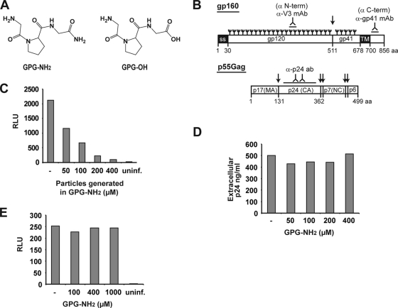
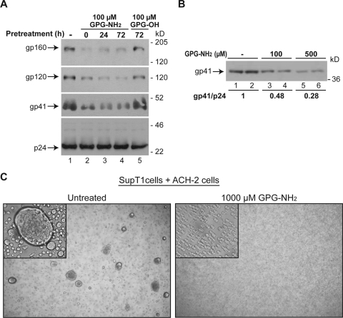
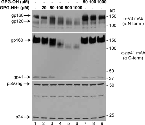
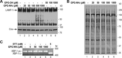
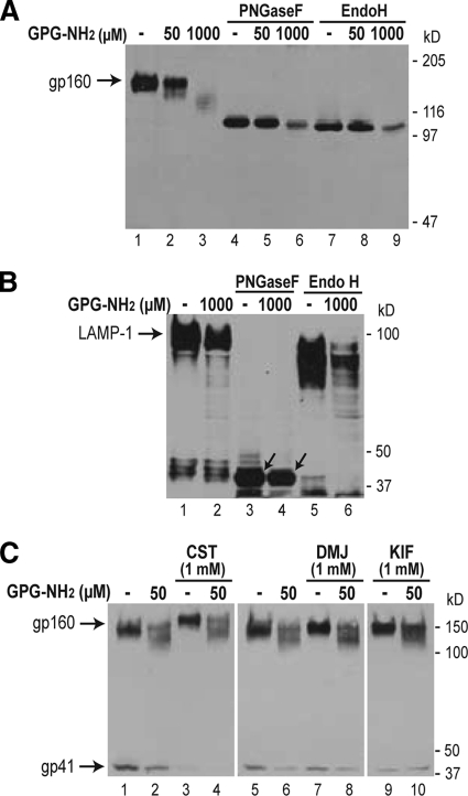
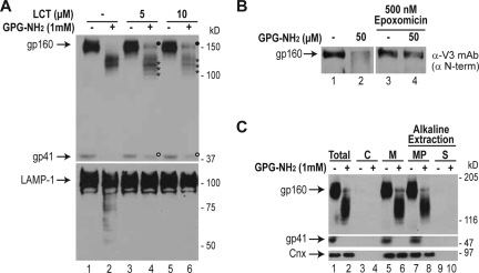
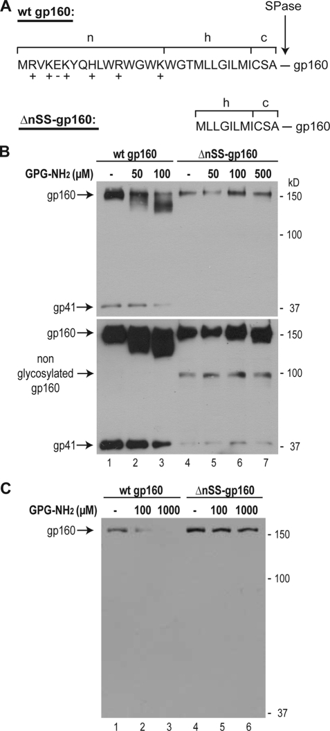
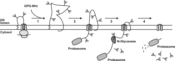
Human immunodeficiency virus type 1 (HIV-1) is dependent on its envelope glycoprotein (Env) to bind, fuse, and subsequently infect a cell. We show here that treatment of HIV-1-infected cells with glycyl-prolyl-glycine amide (GPG-NH(2)), dramatically reduced the infectivity of the released viral particles by decreasing their Env incorporation. The mechanism of GPG-NH(2) was uncovered by examining Env expression and maturation in treated cells. GPG-NH(2) treatment was found to affect Env by significantly decreasing its steady-state levels, its processing into gp120/gp41, and its mass by inducing glycan removal in a manner dependent on its native signal sequence and the proteasome. Therefore, GPG-NH(2) negatively impacts Env maturation, facilitating its targeting for endoplasmic reticulum-associated protein degradation, where Env is deglycosylated en route to its degradation. These findings illustrate that nontoxic drugs such as GPG-NH(2), which can selectively target glycoproteins to existing cellular degradation pathways, may be useful for pathogen therapy.


Figures

FIG. 1.
FIG. 1.
GPG-NH2 treatment reduces HIV-1 particle infectivity. (A) Structure of GPG-NH2 and GPG-OH. (B) Linear structures of the HIV-1 Env precursor protein (gp160) and the precursor protein p55Gag depicting antibody recognition sites (inverted “Y”), N-linked glycans (trident symbol), and proteolytic cleavage sites (↓). (C) HIV-1 particles were produced in the indicated concentrations of GPG-NH2. Equivalent p24 amounts of the viral particles were used to infect TZM-bl cells at 4°C for 2 h, the infection medium was removed, and the cells were cultured for 48 h in the presence of 5 μM indinavir. The luciferase activity in the lysates was monitored to determine the relative infectivity of the particles. (D) ACH-2 cells were pretreated with 0 to 400 μM GPG-NH2 for 24 h before HIV-1 replication was induced with PMA for 48 h. HIV-1 particle production was determined by measuring the extracellular p24 by ELISA. (E) HIV-1 virus was mixed with 0 to 1,000 μM GPG-NH2 and incubated with TZM-bl cells for 2 h. Upon removal of the infection media the cells were incubated in the presence of 5 μM indinavir for 48 h. The cells were subsequently harvested, and the infectivity was determined by measuring the intracellular luciferase activity.
FIG. 2.
FIG. 2.
GPG-NH2 reduces incorporation of Env into HIV-1 particles. (A) ACH-2 cells were pretreated with GPG-NH2 or GPG-OH for the indicated times before induction with PMA for 72 h. The particles were isolated from the extracellular medium by polyethylene glycol precipitation, lysed, separated by SDS-PAGE, immunoblotted, and probed with the α-V3 monoclonal antibody (which recognizes gp120 and gp160), the α-gp41 monoclonal antibody (which recognizes gp41 and gp160), and p24 antiserum. (B) Immunoblot showing the amount of gp41 present in polyethylene glycol-precipitated HIV-1 particles produced by HeLa-tat III cells. The cells pretreated for 20 h with the indicated concentrations of GPG-NH2 prior to transfection with the infectious clone, pNL3-4. The viral particles were isolated 72 h posttransfection and standardized to the extracellular p24 concentrations measured by ELISA. The gp41/p24 ratio was calculated by densitometry. (C) Light microscopy photos of syncytium formation between ACH-2 and CD4-positive SupT1 cells with insets showing higher magnification images. The ACH-2 cells were pretreated for 24 h with 10 μM indinavir and the indicated concentrations of GPG-NH2 prior to PMA stimulation. Five hours later the SupT1 cells were added to the ACH-2 cells, which were then cocultured for 40 h.
FIG. 3.
FIG. 3.
GPG-NH2 caused a decrease in gp160 mobility and steady-state levels. HeLa-tat III cells were transfected with plasmids expressing either gp160 or p55Gag and cultured in the indicated concentrations of GPG-NH2 or GPG-OH for 20 h (gp160) or 48 h (p55Gag). After harvesting, the cell lysates were separated by SDS-PAGE and immunoblotted with antibodies to the N terminus of gp160, α-V3 (upper panel); the C terminus of gp160, α-gp41 (middle panel); or p55Gag/p24 (bottom panel).
FIG. 4.
FIG. 4.
GPG-NH2 affects glycoproteins. (A) Immunoblots of HeLa-tat III cells treated for 48 h as indicated and probed with antibodies to LAMP-1 (upper panel) and calnexin (bottom panel). (B) Glycoprotein blot of HeLa-tat III cell lysates collected from cells cultured in the indicated concentrations of GPG-NH2 for 48 h and probed with concanavalin A. (C) RT-PCR analysis of XBP-1 mRNA splicing using RNA templates from HeLa-tat III cells treated for 48 h with the indicated concentrations of GPG-NH2 or for 3 h with the UPR inducer DTT. The unspliced and spliced XBP-1 mRNAs are designated by “us” and “s,” respectively.
FIG. 5.
FIG. 5.
GPG-NH2 decreases the number of glycans on gp160. (A) Immunoblots of cell lysates prepared from HeLa-tat III cells cultured in the indicated concentrations of GPG-NH2, harvested 20 h posttransfection with gp160, and probed with gp41 antibodies. Where indicated, the lysates were subjected to deglycosylation by PNGaseF or Endo H prior to analysis. (B) HeLa-tat III cells cultured for 48 h in the presence of the indicated GPG-NH2 concentration were harvested and subjected to deglycosylation as in panel A, followed by immunoblotting against LAMP-1. Bands representing fully deglycosylated LAMP-1 are highlighted by arrows. (C) gp160-transfected HeLa-tat III cells were cultured in the indicated concentrations of GPG-NH2 and treated with the glucosidase inhibitor CST and the mannosidase inhibitors DMJ and KIF at 8 h posttransfection. The lysates were harvested 10 h later and subjected to immunoblotting against gp41.
FIG. 6.
FIG. 6.
GPG-NH2 requires functional proteasomes to target gp160 for ERAD. (A) gp160-transfected HeLa-tat III cells were cultured in the indicated concentration of GPG-NH2 and treated with the proteasome inhibitor LCT at 12 h posttransfection. The cells were harvested 10 h later, lysed, separated by SDS-PAGE, and immunoblotted against gp41 (upper panel) or LAMP-1 (bottom panel). Note, in the presence of LCT, the fully glycosylated gp160 (•), the processing to gp41 (○), and the stabilized deglycosylated lower-molecular-weight species (*). (B) gp160-transfected HeLa-tat III cells were cultured in the indicated concentrations of GPG-NH2 and treated with the proteasome inhibitor epoxomicin as described in panel A and immunoblotted against the N terminus of gp160 (α-V3). (C) GPG-NH2-treated HeLa-tat III cells expressing gp160 were homogenized at 20 h posttransfection and fractionated. Immunoblots against gp160/gp41 and calnexin performed on equal fractions from the total homogenate (Total) after removal of the nuclei and nondisrupted cells, the cytosol (C), or the homogenate supernatant that remained after removal of the cellular membranes (M) are shown. The cellular membranes were then subjected to alkaline extraction to separate the integral membrane proteins (MP) from the soluble or peripherally attached proteins (S).
FIG. 7.
FIG. 7.
GPG-NH2 depends on the native gp160 signal sequence to target gp160 for ERAD. (A) The amino acid sequence corresponding to the signal sequence from the HIV-1 Env precursor protein gp160 containing its native signal sequence (wild-type gp160) and the truncated signal sequence (ΔnSS-gp160). The hydrophilic (n), hydrophobic (h), and C-terminal (c) regions, as well as the positively (+) and negatively (−) charged residues and the cleavage site for signal peptidase (SPase), are indicated. (B) Immunoblot of cell lysates from HeLa-tat III cells transfected with either wild-type gp160 or ΔnSSgp160, cultured in the indicated concentrations of GPG-NH2, harvested at 20 h posttransfection, and probed with antibodies against gp41. Both a shorter exposure (upper panel) and a longer exposure (bottom panel) of the same immunoblot are shown. (C) Same as for panel B except that 0.3 μg of vector expressing wild-type gp160 and 1 μg of vector expressing ΔnSS-gp160 were used for transfection to reach similar expression levels of the respective proteins. wt, wild type.
FIG. 8.
FIG. 8.
Proposed model of how GPG-NH2 targets gp160 to ERAD. Initially, gp160 is cotranslationally translocated into the ER. (Arrow 1) The presence of GPG-NH2 or its metabolites prevents the proper folding of gp160, while its signal sequence remains attached. The improper folding of gp160 causes it to be targeted for ERAD. (Arrow 2) gp160 is then retrotranslocated to the cytoplasm in a proteasome-mediated fashion. The polypeptide remains integrated into the ER membrane, likely through its transmembrane region, while (arrow 3) it is deglycosylated by the cytosolic N-glycanase prior to (arrow 4) degradation by the proteasome.

Similar articles

Cited by

References

    1. Abacioglu, Y. H., T. R. Fouts, J. D. Laman, E. Claassen, S. H. Pincus, J. P. Moore, C. A. Roby, R. Kamin-Lewis, and G. K. Lewis. 1994. Epitope mapping and topology of baculovirus-expressed HIV-1 gp160 determined with a panel of murine monoclonal antibodies. AIDS Res. Hum. Retrovir. 10:371-381. - PubMed
    1. Abdurahman, S., A. Vegvari, M. Levi, S. Hoglund, M. Hogberg, W. Tong, I. Romero, J. Balzarini, and A. Vahlne. 2009. Isolation and characterization of a small antiretroviral molecule affecting HIV-1 capsid morphology. Retrovirology 6:34. - PMC - PubMed
    1. Adachi, A., H. E. Gendelman, S. Koenig, T. Folks, R. Willey, A. Rabson, and M. A. Martin. 1986. Production of acquired immunodeficiency syndrome-associated retrovirus in human and nonhuman cells transfected with an infectious molecular clone. J. Virol. 59:284-291. - PMC - PubMed
    1. Ahner, A., and J. L. Brodsky. 2004. Checkpoints in ER-associated degradation: excuse me, which way to the proteasome? Trends Cell Biol. 14:474-478. - PubMed
    1. Andersson, E., P. Horal, A. Jejcic, S. Hoglund, J. Balzarini, A. Vahlne, and B. Svennerholm. 2005. Glycine-amide is an active metabolite of the antiretroviral tripeptide glycyl-prolyl-glycine-amide. Antimicrob. Agents Chemother. 49:40-44. - PMC - PubMed

Publication types