. 2009 Oct;83(19):10211-23.
doi: 10.1128/JVI.00571-09. Epub 2009 Jul 29.

Stable docking of neutralizing human immunodeficiency virus type 1 gp41 membrane-proximal external region monoclonal antibodies 2F5 and 4E10 is dependent on the membrane immersion depth of their epitope regions

Affiliations

Stable docking of neutralizing human immunodeficiency virus type 1 gp41 membrane-proximal external region monoclonal antibodies 2F5 and 4E10 is dependent on the membrane immersion depth of their epitope regions

S Moses Dennison et al. J Virol. 2009 Oct.

Abstract

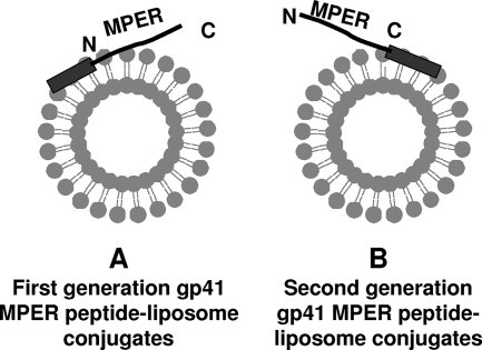
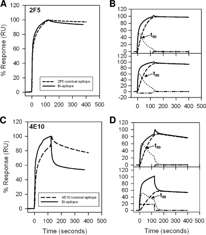
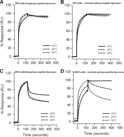
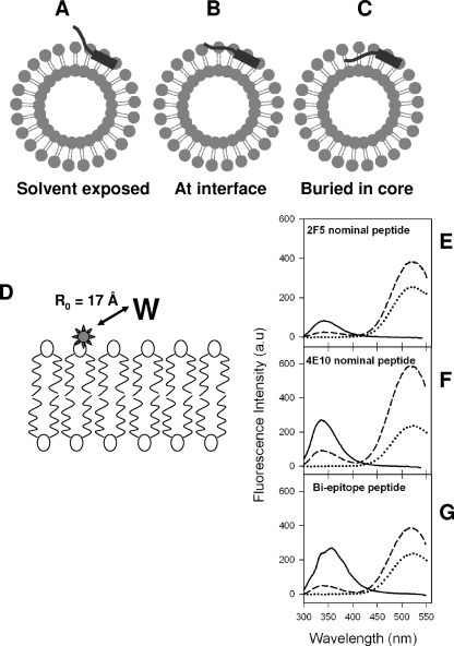
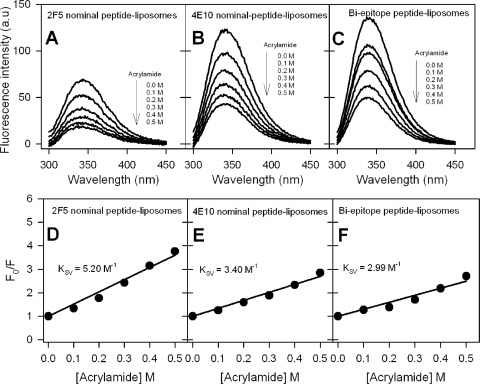
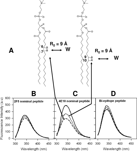
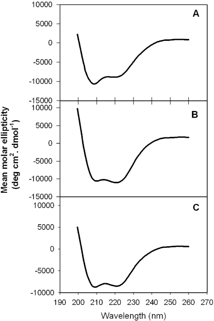
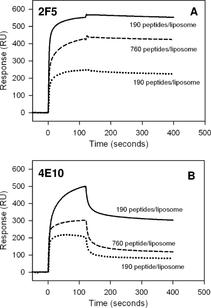
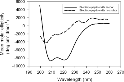
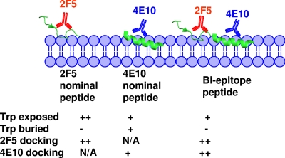
The binding of neutralizing antibodies 2F5 and 4E10 to human immunodeficiency virus type 1 (HIV-1) gp41 involves both the viral membrane and gp41 membrane proximal external region (MPER) epitopes. In this study, we have used several biophysical tools to examine the secondary structure, orientation, and depth of immersion of gp41 MPER peptides in liposomes and to determine how the orientation of the MPER with lipids affects the binding kinetics of monoclonal antibodies (MAbs) 2F5 and 4E10. The binding of 2F5 and 4E10 both to their respective nominal epitopes and to a biepitope (includes 2F5 and 4E10 epitopes) MPER peptide-liposome conjugate was best described by a two-step encounter-docking model. Analysis of the binding kinetics and the effect of temperature on the binding stability of 2F5 and 4E10 to MPER peptide-liposome conjugates revealed that the docking of 4E10 was relatively slower and thermodynamically less favorable. The results of fluorescence-quenching and fluorescence resonance energy transfer experiments showed that the 2F5 epitope was more solvent exposed, whereas the 4E10 epitope was immersed in the polar-apolar interfacial region of the lipid bilayer. A circular dichroism spectroscopic study demonstrated that the nominal epitope and biepitope MPER peptides adopted ordered structures with differing helical contents when anchored to liposomes. Furthermore, anchoring of MPER peptides to the membrane via a hydrophobic anchor sequence was required for efficient MAb docking. These results support the model that the ability of 2F5 and 4E10 to bind to membrane lipid is required for stable docking to membrane-embedded MPER residues. These data have important implications for the design and use of peptide-liposome conjugates as immunogens for the induction of MPER-neutralizing antibodies.


Figures

FIG. 1.
FIG. 1.
Schematic representation of two different strategies of membrane anchoring of MPER peptides. (A) Nonphysiologic anchoring at the amino terminus of MPER used in our earlier report (2). (B) Physiologic anchoring at the carboxyl terminus of MPER used in this study. The hydrophobic membrane-anchoring sequence is shown as a rectangle and was attached at either the amino or carboxyl terminus of the MPER peptide, which is shown as a black line.
FIG. 2.
FIG. 2.
Interaction of MAbs 2F5 and 4E10 with MPER peptide-liposome conjugates. (A) Results for binding of MAb 2F5 to 2F5 nominal epitope peptide-liposome conjugate and biepitope peptide-liposome conjugate are shown. RU, resonance unit. (B) The binding of MAb 2F5 to 2F5 nominal epitope (top) and biepitope (bottom) peptide-liposome conjugates follows the two-step conformational change model. (C and D) Binding of MAb 4E10 to the 4E10 nominal epitope and biepitope peptide-liposome conjugates as described for panels A and B. In each of the overlays (B and D), the binding data are shown by solid lines and represent the observed total binding response. The component curves for the encounter (dotted lines) and docked complexes (dashed lines) were simulated from the experimentally determined rate constants (Table 1). t50 is the time required for half of the encounter complex to be converted to docked complex.
FIG. 3.
FIG. 3.
Temperature dependence of 2F5 and 4E10 binding to peptide-liposome conjugates. Binding kinetics of 2F5 (A and B) and 4E10 (C and D) were measured at different temperatures, as indicated, for the biepitope (A and C) and 2F5 and 4E10 nominal epitope (B and D) peptide-lipid conjugates. The specific binding signal was recorded with reference to the signal for peptide-free synthetic liposomes, as described in Materials and Methods. The data recorded at different temperatures were normalized and are presented as the percent response. RU, relative units.
FIG. 4.
FIG. 4.
Possible orientations of MPER peptides and membrane proximity of tryptophan residues of MPER peptides. (A to C) Pictorial representations showing possible orientations that the MPER peptides could assume when conjugated to liposomes. (D) Schematic diagram showing the location of DANSYL label (star) in the lipid bilayer. The Forster distance (R0) for observing 50% FRET efficiency for the DANSYL-tryptophan pair is indicated (32). (E to G) Tryptophan-to-DANSYL FRET for 2F5 nominal epitope (E), 4E10 nominal epitope (F), and biepitope (G) MPER peptide-liposome conjugates. In each panel, the solid curves show the fluorescence spectra of the peptide-liposome conjugates in the absence of DANSYL-PE, the dashed curves show the fluorescence spectra of peptide-liposome conjugates having 5mol% DANSYL-PE, and the dotted curves show the fluorescence of DANSYL-PE liposomes with no peptides.
FIG. 5.
FIG. 5.
Evaluation of exposure of membrane-anchored 2F5 and 4E10 nominal epitope and biepitope peptides to aqueous quencher. (A to C) Tryptophan fluorescence emission spectra of 2F5 nominal epitope (A), 4E10 nominal epitope (B), and biepitope (C) peptide-liposome conjugates at different added acrylamide concentrations. a.u., arbitrary unit. (D to F) Stern-Volmer plots for the quenching experiments whose results are shown in panels A to C. Ratio of tryptophan fluorescence intensity of peptide-liposomes in the absence of acrylamide to that at different acrylamide concentrations (F0/F) as a function of acrylamide concentration.
FIG. 6.
FIG. 6.
Evaluation of membrane insertion of 2F5 and 4E10 nominal epitope and biepitope peptides. (A) Chemical structures of DBr lipids used in the experiment. The positions of DBr labels and Forster distance (R0) requirements for observing 50% quenching efficiency (5) are also shown. (B to D) Tryptophan fluorescence spectra of 2F5 nominal epitope (B), 4E10 nominal epitope (C), and biepitope (D) peptide-liposome conjugates. In each panel, the solid curves show the spectra recorded with no DBr lipids and the dotted and dashed curves show the spectra recorded with 10 mol% 67DBrPC and 910DBrPC liposomes, respectively. a.u., arbitrary unit.
FIG. 7.
FIG. 7.
CD spectra of MPER peptides conjugated to small unilamellar liposomes. Smoothed CD spectra (see Materials and Methods) are shown for 2F5 nominal epitope (A), 4E10 nominal epitope (B), and biepitope (C) MPER peptide-liposomes. The P/L ratio was kept at 1:200.
FIG. 8.
FIG. 8.
Comparison of 2F5 and 4E10 binding to biepitope MPER peptide presented differently on liposomes. Binding of 2F5 (A) and 4E10 (B) to membrane-anchored biepitope MPER peptide (solid lines) at a P/L ratio of 1:400 and membrane-bound biepitope MPER peptide at P/L ratios of 1:400 (dotted lines) and 1:100 (dashed lines). The surface concentrations of peptides are also indicated. RU, resonance unit.
FIG. 9.
FIG. 9.
CD spectra of biepitope MPER peptides presented differently on small unilamellar liposomes. Smoothed CD spectra of membrane-anchored and membrane-bound biepitope MPER peptides at a P/L ratio of 1:200.
FIG. 10.
FIG. 10.
Pictorial representation of differential membrane immersion of nominal epitope and biepitope MPER peptides influencing the efficiency of 2F5 and 4E10 docking. The 2F5 and 4E10 binding regions of MPER are rendered from their respective Fab-bound crystal structures (6, 25). The different immersion depths indicated for nominal epitope and biepitope peptides in the lipid bilayer were inferred from the fluorescence-quenching and FRET experimental results. The relative stabilities of 2F5 and 4E10 docking shown were derived from the temperature dependence of antibody binding to the respective nominal epitope and biepitope peptide-liposome conjugates. N/A, not applicable. Trp-exposed row, ++ and + indicate a KSV value of 5.0 and ∼3.0 M−1, respectively, in acrylamide quenching experiment. Trp-burried row, + and − represent a ≥25% and <25% quenching of tryptophan fluorescence, respectively, by dibromo lipids. Rows of 2F5 and 4E10 docking, ++ and + represent no change and a twofold change, respectively, in %ΔG (ΔG2/ΔG) on increasing temperature from 10 to 30°C.

Similar articles

Cited by

References

    1. Alam, S. M., S. Dennison, M. Morelli, H. Liao, R. Zhang, B. F. Haynes, S. C. Harrison, and B. Chen. 2008. The role of lipid reactivity of HIV-1 gp41 membrane proximal antibodies in neutralization of HIV-1: novel liposome based immunogen design, abstr. OA07 to 06. Abstracts from AIDS Vaccine 2008, Cape Town, South Africa. October 13-16, 2008. AIDS Res. Hum Retrovir. 24(Suppl. 1):23-24.
    1. Alam, S. M., M. McAdams, D. Boren, M. Rak, R. M. Scearce, F. Gao, Z. T. Camacho, D. Gewirth, G. Kelsoe, P. Chen, and B. F. Haynes. 2007. The role of antibody polyspecificity and lipid reactivity in binding of broadly neutralizing anti-HIV-1 envelope human monoclonal antibodies 2F5 and 4E10 to glycoprotein 41 membrane proximal envelope epitopes. J. Immunol. 178:4424-4435. - PMC - PubMed
    1. Andrade, M. A., P. Chacon, J. J. Merelo, and F. Moran. 1993. Evaluation of secondary structure of proteins from UV circular dichroism spectra using an unsupervised learning neural network. Protein Eng. 6:383-390. - PubMed
    1. Barenholz, Y., D. Gibbes, B. J. Litman, J. Goll, T. E. Thompson, and R. D. Carlson. 1977. A simple method for the preparation of homogeneous phospholipid vesicles. Biochemistry 16:2806-2810. - PubMed
    1. Bolen, E. J., and P. W. Holloway. 1990. Quenching of tryptophan fluorescence by brominated phospholipid. Biochemistry 29:9638-9643. - PubMed

Publication types

LinkOut - more resources