. 2009 Oct;83(19):10299-304.
doi: 10.1128/JVI.00217-09. Epub 2009 Jul 29.

Mapping of functional domains in herpesvirus saimiri complement control protein homolog: complement control protein domain 2 is the smallest structural unit displaying cofactor and decay-accelerating activities

Affiliations

Mapping of functional domains in herpesvirus saimiri complement control protein homolog: complement control protein domain 2 is the smallest structural unit displaying cofactor and decay-accelerating activities

Akhilesh K Singh et al. J Virol. 2009 Oct.

Abstract

Herpesvirus saimiri encodes a functional homolog of human regulator-of-complement-activation proteins named CCPH that inactivates complement by accelerating the decay of C3 convertases and by serving as a cofactor in factor I-mediated inactivation of their subunits C3b and C4b. Here, we map the functional domains of CCPH. We demonstrate that short consensus repeat 2 (SCR2) is the minimum domain essential for classical/lectin pathway C3 convertase decay-accelerating activity as well as for factor I cofactor activity for C3b and C4b. Thus, CCPH is the first example wherein a single SCR domain has been shown to display complement regulatory functions.


Figures

FIG. 1.
FIG. 1.
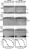
Schematic illustration of sCCPH and SDS-PAGE analysis of purified recombinant sCCPH and its deletion mutants. (Top) Schematic representation of the structure of the soluble form of CCPH (sCCPH), which is composed of four SCRs. The domains are numbered, and the minimum domains shown to be important for C3b and C4b cofactor activities (CFA) and CP DAA are identified. (Bottom) Expressed and purified sCCPH and its deletion mutants were analyzed by 12% (left) and 13% (right) SDS-PAGE under reducing conditions and stained with Coomassie blue. Molecular weights as determined by SDS-PAGE: for sCCPH, 32,000; for SCR1-3, 26,000; for SCR2-4, 27,500; for SCR1-2, 17,000; for SCR2-3, 17,500; for SCR3-4, 16,500; for SCR1, 9,500; for SCR2, 7,000; for SCR3, 8,000; and for SCR4, 8,000. Molecular mass is expressed as kilodaltons in the figure.
FIG. 2.
FIG. 2.
Analysis of factor I cofactor activity of sCCPH and its deletion mutants for human complement proteins C3b and C4b. Cofactor activity was assessed by incubating 3.0 μg of human C3b (upper panels) or C4b (lower panels) with sCCPH/SCR1-3/SCR2-4 (4.0 μM) or SCR1-2/2-3/3-4 (24 μM) in the presence or absence of factor I (100 ng) for the indicated time periods at 37°C in 10 mM sodium phosphate, pH 7.4, containing 145 mM NaCl. The reactions were stopped by addition of sample buffer containing dithiothreitol, and the amount of C3b or C4b cleaved was visualized by subjecting the samples to SDS-PAGE analysis on 10% or 11.5% gel, respectively, and staining with Coomassie blue. During C3b cleavage, the α′-chain is cleaved into N-terminal 68-kDa and C-terminal 46-kDa fragments. The 46-kDa fragment is then cleaved into a 43-kDa fragment. These cleavages indicate inactivation of C3b. In the case of C4b, the α′-chain is cleaved into N-terminal 27-kDa, C-terminal 16-kDa (not visible in the gel), and central C4d fragments. These cleavages indicate the inactivation of C4b.
FIG. 3.
FIG. 3.
Analysis of factor I cofactor activity (CFA) of single SCR mutants of sCCPH for human complement proteins C3b and C4b. (Upper panels) Cofactor activity was assessed by incubating 3.0 μg of human C3b or C4b with the single SCR mutants (44 μM) in the presence or absence of factor I (100 ng) for 4 h at 37°C in PBS (10 mM sodium phosphate, pH 7.4, containing 145 mM NaCl). The reactions were stopped by addition of sample buffer containing dithiothreitol, and the amount of C3b or C4b cleaved was visualized by subjecting the samples to 13% SDS-PAGE and stained with Coomassie blue. Cleavage of the α′-chain of C3b and C4b and generation of cleavage products indicate the inactivation of these proteins. (Middle panels) Human C3b (3.0 μg) or C4b (3.0 μg) and factor I (100 ng) were incubated in PBS with increasing concentrations of sCCPH or the SCR2 mutant at 37°C for 1 h, and the cleavage products were analyzed as described above. (Lower panels) The intensity of the α′-chains of C3b and C4b in the middle panels was determined densitometrically and is represented graphically. The closed and open circles represent sCCPH and the SCR2 mutant, respectively.
FIG. 4.
FIG. 4.
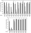
Analysis of CP and AP C3 convertase DAAs of sCCPH and its mutants. (Upper panel) The CP C3 convertase C4b,2a was formed on antibody-coated sheep erythrocytes (EA) by sequentially incubating them with human C1, C4, and C2 (Calbiochem). The C3 convertase on the cells was then allowed to decay by incubating EA-C4b,2a with various concentrations of sCCPH or its mutants for 5 min at 22°C, and the activity of the remaining enzyme was assessed by measuring the cell lysis following incubation for 30 min at 37°C with Guinea pig sera containing 40 mM EDTA (27, 32). (Lower panel) The AP C3 convertase C3b,Bb was formed on sheep erythrocytes (ES) by incubating them with human C3 (Calbiochem) and factors B and D in the presence of NiCl2. The C3 convertase on the cells was then allowed to decay by incubating ES-C3b,Bb with various concentrations of sCCPH or its mutants for 10 min at 37°C, and the activity of the remaining enzyme was assessed by measuring the cell lysis following incubation with EDTA-sera for 30 min at 37°C (35, 37). The data obtained were normalized by considering the lysis that occurred in the absence of an inhibitor as 100% lysis.
FIG. 5.
FIG. 5.
Binding of sCCPH and its mutants to C3b and C4b. Binding was determined by a surface plasmon resonance-based assay (38). Sensograms were generated by immobilizing biotinylated C3b (1,200 response units [RUs]) and C4b (940 RUs) on streptavidin chips (Sensor Chip SA; Biacore AB; additional RUs of C3b [∼6,000 RUs] were deposited by forming AP C3 convertase on the chip and flowing native C3 [14]) and injecting sCCPH or its mutants in PBS-T (10 mM sodium phosphate and 145 mM NaCl, pH 7.4, containing 0.05% Tween 20) over the chip. Flow cells immobilized with bovine serum albumin-biotin (Sigma) served as control flow cells. (Left panels) Binding of sCCPH and its various mutants to C3b (top) and C4b (bottom). The sensograms were generated by injecting 500 nM and 2 μM of sCCPH and its various mutants over C3b and C4b chips, respectively. (Middle panels) Sensogram overlay for the interaction between sCCPH and C3b (top) or sCCPH and C4b (bottom). (Right panels) Sensogram overlay for the interaction between SCR2-4 and C3b (top) and SCR1-3 and C4b (bottom). The concentrations of proteins injected are indicated at the right of the sensograms. The solid lines in the top middle and top right panels represent the global fitting of the data to a 1:1 Langmuir binding model with a drifting baseline (A + B ↔ AB; Biaevaluation 4.1). The small arrows in the bottom middle and right panels indicate the time points used for evaluating the steady-state affinity data.

Similar articles

Cited by

References

    1. Ahmad, M., K. Pyaram, J. Mullick, and A. Sahu. 2007. Viral complement regulators: the expert mimicking swindlers. Indian J. Biochem. Biophys. 44:331-343. - PubMed
    1. Albrecht, J. C., and B. Fleckenstein. 1992. New member of the multigene family of complement control proteins in herpesvirus saimiri. J. Virol. 66:3937-3940. - PMC - PubMed
    1. Blom, A. M., L. Kask, and B. Dahlback. 2001. Structural requirements for the complement regulatory activities of C4BP. J. Biol. Chem. 276:27136-27144. - PubMed
    1. Bramley, J. C., A. Davies, and P. J. Lachmann. 1997. Herpesvirus saimiri CD59—baculovirus expression and characterisation of complement inhibitory activity. Biochem. Soc. Trans. 25:354S. - PubMed
    1. Brodbeck, W. G., D. Liu, J. Sperry, C. Mold, and M. E. Medof. 1996. Localization of classical and alternative pathway regulatory activity within the decay-accelerating factor. J. Immunol. 156:2528-2533. - PubMed

Publication types

LinkOut - more resources