. 2009 Oct;83(19):10007-15.
doi: 10.1128/JVI.00559-09. Epub 2009 Jul 29.

Latency-associated transcript (LAT) exon 1 controls herpes simplex virus species-specific phenotypes: reactivation in the guinea pig genital model and neuron subtype-specific latent expression of LAT

Affiliations

Latency-associated transcript (LAT) exon 1 controls herpes simplex virus species-specific phenotypes: reactivation in the guinea pig genital model and neuron subtype-specific latent expression of LAT

Andrea S Bertke et al. J Virol. 2009 Oct.

Abstract

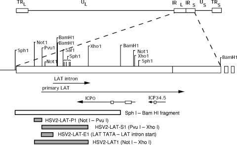
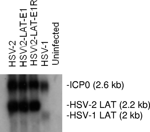
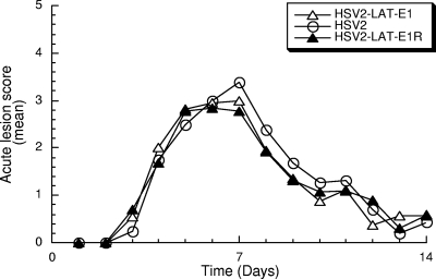
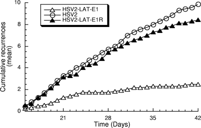
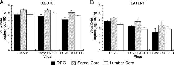
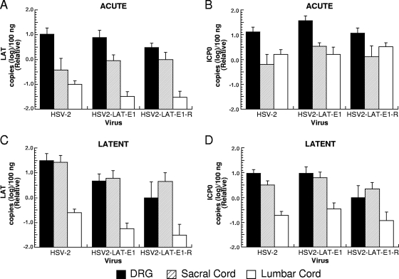
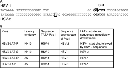
Herpes simplex virus 1 (HSV-1) and HSV-2 cause similar acute infections but differ in their abilities to reactivate from trigeminal and lumbosacral dorsal root ganglia. During latency, HSV-1 and HSV-2 also preferentially express their latency-associated transcripts (LATs) in different sensory neuronal subtypes that are positive for A5 and KH10 markers, respectively. Chimeric virus studies showed that LAT region sequences influence both of these viral species-specific phenotypes. To further map the LAT region sequences responsible for these phenotypes, we constructed the chimeric virus HSV2-LAT-E1, in which exon 1 (from the LAT TATA to the intron splice site) was replaced by the corresponding sequence from HSV-1 LAT. In intravaginally infected guinea pigs, HSV2-LAT-E1 reactivated inefficiently relative to the efficiency of its rescuant and wild-type HSV-2, but it yielded similar levels of viral DNA, LAT, and ICP0 during acute and latent infection. HSV2-LAT-E1 preferentially expressed the LAT in A5+ neurons (as does HSV-1), while the chimeric viruses HSV2-LAT-P1 (LAT promoter swap) and HSV2-LAT-S1 (LAT sequence swap downstream of the promoter) exhibited neuron subtype-specific latent LAT expression phenotypes more similar to that of HSV-2 than that of HSV-1. Rescuant viruses displayed the wild-type HSV-2 phenotypes of efficient reactivation in the guinea pig genital model and a tendency to express LAT in KH10+ neurons. The region that is critical for HSV species-specific differences in latency and reactivation thus lies between the LAT TATA and the intron splice site, and minor differences in the 5' ends of chimeric sequences in HSV2-LAT-E1 and HSV2-LAT-S1 point to sequences immediately downstream of the LAT TATA.


Figures

FIG. 1.
FIG. 1.
Virus construction. Genomic and endonuclease restriction sites are shown relative to HSV-2 strain HG52. The HSV genome consists of unique long and short (UL and US) regions flanked by internal and terminal repeats (IRL/IRS and TRL/TRS). The primary LAT is transcribed from the repeat regions in the direction antisense to ICP0 and ICP34.5 (transcripts are shown here as lines, with arrows indicating the direction of transcription). The primary LAT is spliced into a single stable LAT intron in HSV-2 and two differentially spliced introns in HSV-1. Boxes on the ICP0 and ICP34.5 lines denote introns that are not readily detectable after splicing. Chimeric viruses (HSV2-LAT-P1, HSV2-LAT-S1, HSV2-LAT-E1, and HSV2-LAT1) were constructed by homologous recombination into wild-type virus after cloning HSV-1 sequences corresponding to the shaded areas into the SphI-BamHI clone. The location of the PvuI site relative to the LAT TATAs is illustrated in Fig. 6.
FIG. 2.
FIG. 2.
Northern hybridization of RNA from Vero cells 16 h after infection with HSV-2, HSV2-LAT-E1, HSV2-LAT-E1R, and HSV-1. RNA was probed with an SalI-XhoI probe overlapping the HSV-2 LAT intron, ICP0, with partial homology to these RNAs from HSV-1. Expected sizes of LAT and ICP0 are shown.
FIG. 3.
FIG. 3.
Acute lesion severity. Lesion severity (as measured on a 4-point scale) is graphed as the mean for each group of guinea pigs on each day of observation from days 1 to 14 p.i. Viruses included HSV-2 (n = 19), HSV2-LAT-E1 (n = 25), and HSV2-LAT-E1R (n = 13). There were no significant differences among the areas under the curve for each virus (P = 0.536 by Kruskal-Wallis test).
FIG. 4.
FIG. 4.
Cumulative recurrences per guinea pig for each group during latent infection. Viruses tested included HSV-2 (n = 7), HSV2-LAT-E1 (n = 13), and HSV2-LAT-E1R (n = 7). Guinea pigs that did not survive acute disease were excluded. P values (using the Mann-Whitney test) for pairwise comparisons of recurrence frequency are the following: for HSV-2 versus HSV2-LAT-E1, P < 0.0005; for HSV2-LAT-E1 versus HSV2-LAT-E1R, P = 0.001; and for HSV-2 versus HSV2-LAT-E1R, P = 0.221.
FIG. 5.
FIG. 5.
Viral DNA quantities of HSV-2, HSV2-LAT-E1, and HSV2-LAT-E1R in the DRG, sacral cord, and lumbar cord during acute and latent infection. Viral DNA extracted from the lumbosacral DRG, sacral spinal cord, and lumbar spinal cord during acute infection (day 8 p.i.) (A) and latent infection (day 42 p.i.) (B) was quantified by TaqMan PCR assay and normalized to the gene for the 18S rRNA. The day 8 experiment included HSV-2 (n = 10), HSV2-LAT-E1 (n = 11), and HSV2-LAT-E1R (5). The day 42 experiment included HSV-2 (n = 9), HSV2-LAT-E1 (n = 14), and HSV2-LAT-E1R (n = 8).
FIG. 6.
FIG. 6.
LAT and ICP0 transcript expression in DRG, sacral spinal cord, and lumbar spinal cord during acute and latent infection (RNA copies [log]/100 ng total RNA). RNA extracted from the lumbosacral DRG, sacral spinal cord, and lumbar spinal cord during acute infection (day 8 p.i.) (A and B) and latent infection (day 42 p.i.) (C and D) was quantified by TaqMan PCR assay with primers and probes specific for HSV-2 LAT (A and C) and ICP0 (B and D). 18S rRNA also was quantified by TaqMan RT-PCR and used to normalize for RNA loading. Results are shown relative to mean quantities of each transcript that were detected in lumbosacral DRG infected with HSV2-LAT-E1R on day 42. Viruses tested at day 8 included HSV2-LAT-E1 (n = 11), HSV-2 (n = 10), and HSV2-LAT-E1R (n = 5). Viruses tested at day 42 included HSV2-LAT-E1 (n = 14), HSV-2 (n = 9), and HSV2-LAT-E1R (n = 8).
FIG. 7.
FIG. 7.
Sequences and phenotypes. (A) Comparison of HSV-1 and HSV-2 sequences between LAT TATAs and PvuI recognition sites. Chimeric viruses HSV2-LAT-P1 and HSV2-LAT-E1 contain HSV-1 sequences in this region, while HSV2-LAT-S1 contains HSV-2 sequences in this region. Potential binding sites for ICP4 and SP1 are shown. An EGR-1 consensus binding sequence (GCGGGGGCG) is italicized in the HSV-1 sequence. The PvuI sites are displayed in boldface. Previously mapped start sites for HSV-1 and HSV-2 primary LATs are shown in boxes. (B) Correlation of sequences near the LAT 5′ end with the chimeric virus latency establishment phenotype.

References

    1. Batchelor, A. H., K. W. Wilcox, and P. O'Hare. 1994. Binding and repression of the latency-associated promoter of herpes simplex virus by the immediate early 175K protein. J. Gen. Virol. 75:753-767. - PubMed
    1. Berthomme, H., J. Lokensgard, L. Yang, T. Margolis, and L. T. Feldman. 2000. Evidence for a bidirectional element located downstream from the herpes simplex virus type 1 latency-associated promoter that increases its activity during latency. J. Virol. 74:3613-3622. - PMC - PubMed
    1. Bertke, A. S., A. Patel, and P. R. Krause. 2007. Herpes simplex virus latency-associated transcript sequence downstream of the promoter influences type-specific reactivation and viral neurotropism. J. Virol. 81:6605-6613. - PMC - PubMed
    1. Bhattacharjee, P. S., R. K. Tran, M. E. Myles, K. Maruyama, A. Mallakin, D. C. Bloom, and J. M. Hill. 2003. Overlapping subdeletions within a 348-bp in the 5′ exon of the LAT region that facilitates epinephrine-induced reactivation of HSV-1 in the rabbit ocular model do not further define a functional element. Virology 312:151-158. - PubMed
    1. Bloom, D. C., G. B. Devi-Rao, J. M. Hill, J. G. Stevens, and E. K. Wagner. 1994. Molecular analysis of herpes simplex virus type 1 during epinephrine-induced reactivation of latently infected rabbits in vivo. J. Virol. 68:1283-1292. - PMC - PubMed

Substances