. 2009 Sep;151(1):78-87.
doi: 10.1104/pp.109.140582. Epub 2009 Jul 29.

Mutations in UDP-Glucose:sterol glucosyltransferase in Arabidopsis cause transparent testa phenotype and suberization defect in seeds

Affiliations

Mutations in UDP-Glucose:sterol glucosyltransferase in Arabidopsis cause transparent testa phenotype and suberization defect in seeds

Seth DeBolt et al. Plant Physiol. 2009 Sep.

Abstract

In higher plants, the most abundant sterol derivatives are steryl glycosides (SGs) and acyl SGs. Arabidopsis (Arabidopsis thaliana) contains two genes, UGT80A2 and UGT80B1, that encode UDP-Glc:sterol glycosyltransferases, enzymes that catalyze the synthesis of SGs. Lines having mutations in UGT80A2, UGT80B1, or both UGT80A2 and UGT8B1 were identified and characterized. The ugt80A2 lines were viable and exhibited relatively minor effects on plant growth. Conversely, ugt80B1 mutants displayed an array of phenotypes that were pronounced in the embryo and seed. Most notable was the finding that ugt80B1 was allelic to transparent testa15 and displayed a transparent testa phenotype and a reduction in seed size. In addition to the role of UGT80B1 in the deposition of flavanoids, a loss of suberization of the seed was apparent in ugt80B1 by the lack of autofluorescence at the hilum region. Moreover, in ugt80B1, scanning and transmission electron microscopy reveals that the outer integument of the seed coat lost the electron-dense cuticle layer at its surface and displayed altered cell morphology. Gas chromatography coupled with mass spectrometry of lipid polyester monomers confirmed a drastic decrease in aliphatic suberin and cutin-like polymers that was associated with an inability to limit tetrazolium salt uptake. The findings suggest a membrane function for SGs and acyl SGs in trafficking of lipid polyester precursors. An ancillary observation was that cellulose biosynthesis was unaffected in the double mutant, inconsistent with a predicted role for SGs in priming cellulose synthesis.


Figures
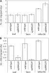
Figure 1.
Figure 1.
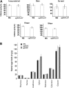
Analysis of sterols and sterol derivates in ugt80A2,B1 mutant relative to wild type. Total sterol derivatives were quantified and compared between wild-type and mutant plants in mg per gram dry weight−1 as FS + SE (A) and SG + ASG (B). FS + SE was the sum of FS measured by GC-FID + sterols measured by GC-FID released from SE fraction after saponification. Values are the mean of three replicates and experimental analysis was duplicated; error bars indicate se from the mean. Inflor/Sili, Inflorescence/silique.
Figure 2.
Figure 2.
Elongation defects in ugt80A2,B1 double-mutant embryos. Nomarski images of embryogenesis during globular (A–C), heart (D and E), torpedo (F and G), bent-cotyledon (H and I), and mature embryo (J) stages. Each vertical section exhibits the identical scale so that the sizes of the mutant embryos can be directly compared with that of wild type (WT; top row). Deviations from the wild-type morphology are first apparent at the late heart stage (F). The ugt80A2,B1 mutant displays elongation defects in outgrowth of the cotyledon primordia (yellow dotted lines). Elongation defects along the apical-basal axis are more obvious in the torpedo, bent-cotyledon, and mature stages. Red dotted lines indicate shorter hypocotyl and root lengths for ugt80A2,B1 at the mature embryo stage (J). Bars = 50 μm. [See online article for color version of this figure.]
Figure 3.
Figure 3.
Cellulose and cell wall analysis. A, Cellulose composition was measured for wild-type WS-O and double mutant in various tissues. B, Cell wall neutral sugar composition of the ugt80A2 and ugt80B1 single mutants was analyzed and compared with that of wild-type WS-O plants.
Figure 4.
Figure 4.
Transparent testa phenotype of ugt80B1 and ugt80A2,B1 mutants. A, Light microscopy to visualize seed coloration in wild type (WT), ugt80A2, ugt80B1, rescue, and ugt80A2,B1. B, Tetrazolium red uptake in wild type, ugt80A2, ugt80B1, rescue, and ugt80A2,B1. C, DMACA staining shows altered flavanol composition in wild type, ugt80A2, ugt80B1, rescue, and ugt80A2,B1 (scale bars = 150 μm).
Figure 5.
Figure 5.
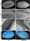
Scanning and transmission electron micrographs and suberin assessment of seed phenotype indicate aberrant lipid polyester production. A and C, Wild-type (WT) seeds exhibit uniform cell shapes and well-formed columella. B and D, Double-mutant seed with a collapsed region of the seed near the funiculus. Columella exhibit aberrant morphology (scale bar in A and B = 100 μm; C and D = 15 μm). E, TEM micrograph of the outer layer of a mature wild-type seed. An electron-dense cuticle covers the outer seed coat. The integuments have collapsed into a dense brown pigment layer. F, Micrograph of double-mutant seed at maturity. The electron-dense cuticle layer is absent (arrow; scale bar = 1 μm). G and H, Suberin autofluorescence in wild-type seeds (G) by illumination under 365-nm UV light compared with the ugt80B1 mutant (H), which lacks suberin accumulation (arrows; scale as in A and B). bpl, Brown pigment layer; ed cyt, electron dense regions in the cytoplasm; en, endosperm; ext, exterior; ow, outer cell wall. [See online article for color version of this figure.]
Figure 6.
Figure 6.
Lipid polyester monomers from seeds of wild-type and ugt80A2,B1 plants. The insoluble dry residue obtained after grinding and delipidation of tissues with organic solvents was depolymerized by acid-catalyzed methanolysis and aliphatic and aromatic monomers released were analyzed by GC-MS. Values are means of four replicates. Error bars denote sds. DCAs, Dicarboxylic acids; FAs, fatty acids; PAs, primary alcohols; br., branched.

References

    1. Abraham W, Wertz PW, Burken RR, Downing DT (1987) Glucosylsterol and acylglucosylsterol of snake epidermis: structure determination. J Lipid Res 28 446–449 - PubMed
    1. Baxter IR, Young JC, Armstrong G, Foster N, Bogenschutz N, Cordova T, Peer WA, Hazen SP, Murphy AS, Harper JF (2005) A plasma membrane H+-ATPase is required for the formation of proanthocyanidins in the seed coat endothelium of Arabidopsis thaliana. Proc Natl Acad Sci USA 102 2649–2654 - PMC - PubMed
    1. Beisson F, Li YH, Bonaventure G, Pollard M, Ohlrogge JB (2007) The acyltransferase GPAT5 is required for the synthesis of suberin in seed coat and root of Arabidopsis. Plant Cell 19 351–368 - PMC - PubMed
    1. Blumenkranz N, Asboe-Hanson G (1973) New methods for quantitative determination of uronic acids. Anal Biochem 54 484–489 - PubMed
    1. Bouvier-Nave P, Ullmann P, Rimmele D, Benveniste P (1984) Phospholipid dependence of plant UDP-glucose sterol β-D-glucosyl transferase. I. Detergent mediated delipidation by selective solubilization. Plant Sci Lett 36 19–27

Publication types

MeSH terms