. 2009 Jul 29;29(30):9429-38.
doi: 10.1523/JNEUROSCI.1472-09.2009.

Dynein light chain LC8 regulates syntaphilin-mediated mitochondrial docking in axons

Affiliations

Dynein light chain LC8 regulates syntaphilin-mediated mitochondrial docking in axons

Yan-Min Chen et al. J Neurosci. .

Abstract

Mitochondria in the cell bodies of neurons are transported down neuronal processes in response to changes in local energy and metabolic states. Because of their extreme polarity, neurons require specialized mechanisms to regulate mitochondrial transport and retention in axons. Our previous studies using syntaphilin (snph) knock-out mice provided evidence that SNPH targets to axonal mitochondria and controls their mobility through its static interaction with microtubules (MTs). However, the mechanisms regulating SNPH-mediated mitochondrial docking remain elusive. Here, we report an unexpected role for dynein light chain LC8. Using proteomic biochemical and cell biological assays combined with time-lapse imaging in live snph wild-type and mutant neurons, we reveal that LC8 regulates axonal mitochondrial mobility by binding to SNPH, thus enhancing the SNPH-MT docking interaction. Using mutagenesis assays, we mapped a seven-residue LC8-binding motif. Through this specific interaction, SNPH recruits LC8 to axonal mitochondria; such colocalization is abolished when neurons express SNPH mutants lacking the LC8-binding motif. Transient LC8 expression reduces mitochondrial mobility in snph (+/+) but not (-/-) neurons, suggesting that the observed effect of LC8 depends on the SNPH-mediated docking mechanism. In contrast, deleting the LC8-binding motif impairs the ability of SNPH to immobilize axonal mitochondria. Furthermore, circular dichroism spectrum analysis shows that LC8 stabilizes an alpha-helical coiled-coil within the MT-binding domain of SNPH against thermal unfolding. Thus, our study provides new mechanistic insights into controlling mitochondrial mobility through a dynamic interaction between the mitochondrial docking receptor and axonal cytoskeleton.


Figures

Figure 1.
Figure 1.
SNPH selectively interacts with LC8 via its seven-residue LC8-binding motif. A, Schematic diagram of SNPH domain structures and truncated mutants used in GST pull-down study. MitoBD, Mitochondrial-binding domain. B, Coomassie blue staining of GST pull-down from rat brain homogenates. GST-SNPH (203–469), but not (1–130) and GST control, pulled down a protein band at 10 kDa (red box), which was then eluted for mass spectrometry and matches the sequences of rat LC8-1 and LC8-2. C, Immunoblot with anti-LC8 antibody confirmed the protein band as LC8. Dynein intermediate chain (IC74) was not detected in the same pull-down assay. D, Immunoprecipitation of SNPH with LC8 from brain homogenates of snph wild-type mouse but not from snph (−/−) mouse brain by an anti-SNPH antibody. IC74 was not detected from the same assays. Preimmune serum (PIS) was used as a control. E, Pull-down assays revealed that the SNPH sequence from residues 282 to 380 is sufficient for stoichiometrical binding to LC8 (red box). F, Immunoprecipitation of GFP-SNPH with HA-LC8 from cotransfected COS7 cells further revealed a seven-residue sequence of SNPH (355–361) as the LC8-binding motif. Deleting this motif abolished its interaction with HA-LC8. MW, Molecular weight; HA, hemagglutinin; IP, immunoprecipitation; IB, immunoblot; BH, brain homogenates; WT, wild type; KO, knock-out.
Figure 2.
Figure 2.
SNPH recruits LC8 to axonal mitochondria via its LC8-binding motif. Representative axonal images of hippocampal neurons cotransfected at DIV9 with GFP-LC8 and DsRed-mito (left), or combined with SNPH (middle) or SNPHΔ355–361 (right). Images were taken 4 d after transfection. The corresponding profiles (bottom panels) were plotted based on fluorescence intensities of GFP-LC8 and DsRed-mito and reflect the relative codistribution of LC8 (green) with mitochondria (red) along axonal processes. LC8, cytosolic protein, is diffused throughout the axonal cytoplasm. Coexpressing SNPH redistributes LC8 to mitochondria. Note that deleting the LC8-binding motif (SNPHΔ355–361) abolishes its role in recruiting GFP-LC8 to axonal mitochondria. a.u., Arbitrary units. Scale bars, 10 μm.
Figure 3.
Figure 3.
Deleting LC8-binding motif reduces the efficiency of SNPH in docking axonal mitochondria. A, B, Representative kymographs showing relative mobility of axonal mitochondria labeled by GFP-SNPH (A) or GFP-SNPHΔ355–361 (B). Neurons were cotransfected at DIV9 with DsRed-mito and GFP-SNPH (A) or GFP-SNPHΔ355–361 (B). Axonal mitochondrial motility was assessed in live neurons 3 d after transfection. Motion data are presented as a kymograph, in which vertical lines represent stationary mitochondria and slanted lines or curves indicate motile ones. Note that all GFP-SNPH-labeled mitochondria remain stationary whereas some of GFP-SNPHΔ355–361-labeled mitochondria are mobile (indicated by white arrows in B). Yellow arrow (in A) points to a motile mitochondrion unlabeled by GFP-SNPH, as a time-lapse imaging control. Scale bars, 20 μm.
Figure 4.
Figure 4.
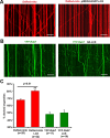
Elevated expression of LC8 selectively inhibits the mobility of axonal mitochondria. A, Representative kymographs showing axonal mitochondrial mobility. Hippocampal neurons were transfected at DIV9 with DsRed-Mito (left) or cotransfected with DsRed-Mito and pIRES2-EGFP-LC8 (right). B, Representative kymographs showing late endosomal mobility in axons of hippocampal neurons transfected at DIV9 with YFP-Rab7 (left) or cotransfected with YFP-Rab7 and HA-LC8 (right). Axonal mitochondrial and late endosomal motility was observed by time-lapse imaging in live neurons 3 d after transfection. Scale bars, 20 μm. C, Relative docked axonal mitochondria (red) and late endosomes (green). Error bars: SEM; “n” indicates the number of axons imaged. HA, Hemagglutinin. **p < 0.01, t test.
Figure 5.
Figure 5.
Role of LC8 in inhibiting mitochondrial mobility depends on SNPH. A, B, Representative kymographs showing axonal mitochondrial mobility in snph (−/−) hippocampal neurons transfected at DIV9 with DsRed-Mito (A) or cotransfected with DsRed-Mito and pIRES2-EGFP-LC8 (B). Neurons were time-lapse imaged 3 d posttransfection. Scale bars, 20 μm.
Figure 6.
Figure 6.
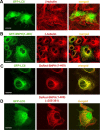
LC8 associates with MTs via its binding to SNPH. A, B, COS7 cells were transfected with GFP-LC8 (A) or GFP-SNPH (1–469) (B) followed by immunostaining with anti-β-tubulin antibody 1 d posttransfection. Note that GFP-LC8 exhibits diffuse distribution in both cytoplasm and nuclei in the absence of exogenously expressed SNPH, whereas GFP-SNPH (1–469) alone predominantly associates with MTs. C, D, COS cells were cotransfected with GFP-LC8 and DsRed-SNPH(1-469) (C) or DsRed-SNPH(1–469)(Δ355–361) lacking the LC8-binding motif (D) followed by imaging 1 d after transfection. Note that deleting the LC-binding motif abolishes the role of SNPH in attaching LC8 to MTs. Scale bars, 20 μm.
Figure 7.
Figure 7.
CD spectrum reveals the role of LC8 in stabilizing the MT-binding domain of SNPH against thermal unfolding. A, Schematic diagram of SNPH segments containing an α-helical coiled-coil (130–203) MTB, and its mutant deleting the LC8-binding motif (355–361) used in far-UV CD measurements. B, CD spectra of purified LC8, SNPH (1–469), and SNPH (110–380) at a concentration of 10 μm in 20 mm phosphate buffer, pH 7.4, containing 150 mm NaCl and 0.5 mm DTT at 25°C. C, Thermal denaturation curves of SNPH (110–380) alone (black ▲) and mixture of SNPH (110–380) and LC8 after the subtraction of LC8 spectrum (red ●). D, Thermal denaturation curves of SNPH(110–380)(Δ355–361) alone (black ▲) and a mixture of SNPH(110–380)(Δ355–361) and LC8 after the subtraction of LC8 spectrum (red ●).
Figure 8.
Figure 8.
LC8 enhances cosedimentation of SNPH with MTs in spin-down assay. GST-SNPH (1–469) or the mixture of GST-SNPH (1–469) and LC8 was incubated in the absence or presence of Taxol-stabilized MTs. Following centrifugation at 100,000 × g for 40 min, the supernatant (S) and pellets (P) were analyzed by SDS-PAGE and visualized by Coomassie blue staining. Note that LC8 remained in the supernatant in the absence of SNPH, whereas a significant part of SNPH (1–469) was spun down with MTs in the absence of LC8. Coincubating SNPH (1–469) and LC8 enhanced cosedimentation of both proteins with MTs (highlighted by red boxes). MW, Molecular weight.
Figure 9.
Figure 9.
Proposed model of LC8 as the “stabilizer” of the SNPH-MT docking interaction. SNPH acts as a receptor for docking/anchoring mitochondria at MTs in axons and is required for maintaining a large number of axonal mitochondria in a stationary state. LC8 binds to SNPH through the conservative LC8-binding motif and serves as the “stabilizer” of the SNPH-coiled-coil structure within the MT-binding domain, thus increasing the docking efficiency.

Similar articles

Cited by

References

    1. Barbar E. Dynein light chain LC8 is a dimerization hub essential in diverse protein networks. Biochemistry. 2008;47:503–508. - PubMed
    1. Barbar E, Kleinman B, Imhoff D, Li M, Hays TS, Hare M. Dimerization and folding of LC8, a highly conserved light chain of cytoplasmic dynein. Biochemistry. 2001;40:1596–1605. - PubMed
    1. Benashski SE, Harrison A, Patel-King RS, King SM. Dimerization of the highly conserved light chain shared by dynein and myosin V. J Biol Chem. 1997;272:20929–20935. - PubMed
    1. Bergen JM, Pun SH. Evaluation of an LC8-binding peptide for the attachment of artificial cargo to dynein. Mol Pharm. 2007;4:119–128. - PMC - PubMed
    1. Billups B, Forsythe ID. Presynaptic mitochondrial calcium sequestration influences transmission at mammalian central synapses. J Neurosci. 2002;22:5840–5847. - PMC - PubMed

Publication types

MeSH terms