. 2009 Jul 29;29(30):9439-49.
doi: 10.1523/JNEUROSCI.6055-08.2009.

Neuronal 3',3,5-triiodothyronine (T3) uptake and behavioral phenotype of mice deficient in Mct8, the neuronal T3 transporter mutated in Allan-Herndon-Dudley syndrome

Affiliations

Neuronal 3',3,5-triiodothyronine (T3) uptake and behavioral phenotype of mice deficient in Mct8, the neuronal T3 transporter mutated in Allan-Herndon-Dudley syndrome

Eva K Wirth et al. J Neurosci. .

Abstract

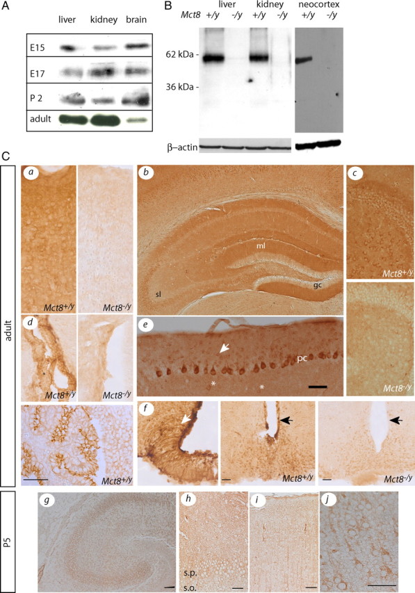
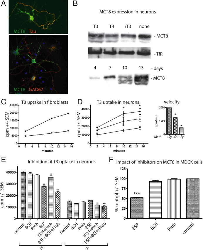
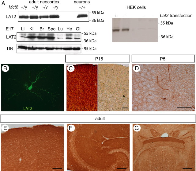
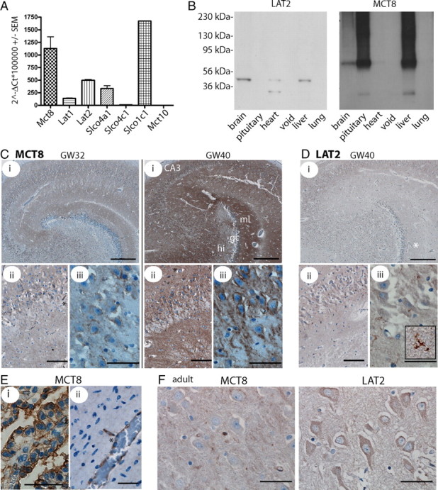
Thyroid hormone transport into cells requires plasma membrane transport proteins. Mutations in one of these, monocarboxylate transporter 8 (MCT8), have been identified as underlying cause for the Allan-Herndon-Dudley syndrome, an X-linked mental retardation in which the patients also present with abnormally high 3',3,5-triiodothyronine (T(3)) plasma levels. Mice deficient in Mct8 replicate the thyroid hormone abnormalities observed in the human condition. However, no neurological deficits have been described in mice lacking Mct8. Therefore, we subjected Mct8-deficient mice to a comprehensive immunohistochemical, neurological, and behavioral screen. Several behavioral abnormalities were found in the mutants. Interestingly, some of these behavioral changes are compatible with hypothyroidism, whereas others rather indicate hyperthyroidism. We thus hypothesized that neurons exclusively dependent on Mct8 are in a hypothyroid state, whereas neurons expressing other T(3) transporters become hyperthyroid, if they are exposed directly to the high plasma T(3). The majority of T(3) uptake in primary cortical neurons is mediated by Mct8, but pharmacological inhibition suggested functional expression of additional T(3) transporter classes. mRNAs encoding six T(3) transporters, including L-type amino acid transporters (LATs), were coexpressed with Mct8 in isolated neurons. We then demonstrated Lat2 expression in cultured neurons and throughout murine brain development. In contrast, LAT2 is expressed in microglia in the developing human brain during gestation, but not in neurons. We suggest that lack of functional complementation by alternative thyroid hormone transporters in developing human neurons precipitates the devastating neurodevelopmental phenotype in MCT8-deficient patients, whereas Mct8-deficient mouse neurons are functionally complemented by other transporters, for possibly Lat2.


Figures

Figure 1.
Figure 1.
Developmental and cell type-specific expression of MCT8 immunoreactivity in the mouse brain. A, Western blots demonstrating the relative abundance of cerebrocortical MCT8 protein compared with liver and kidney at E15, E17, P2, and adult. B, Specificity of the MCT8 antiserum in Western blot. Molecular weight markers are indicated on the left. β-Actin was used as loading control. C, MCT8 immunoreactivity in Mct8 +/y brain compared with Mct8 −/y controls. a, Cerebral cortex. b, Hippocampus. c, Dentate gyrus with hilus. d, Choroid plexus. The bottom panel is from embryonic day 15. e, Cerebellar cortex. Scale bar, 50 μm. f, Hypothalamus. g, Hippocampus at P5. Scale bar, 100 μm. h, CA3 region magnified from g. s.p., Stratum pyramidale; s.o., stratum oriens. i, Somatosensory cortex at P5. j, Magnified view of i. Scale bars: h–j, 50 μm.
Figure 2.
Figure 2.
Immunohistochemical analysis of Mct8-deficient mouse brains. A, Cerebral cortex (S1BF). No differences were observed in the staining patterns for NeuN, parvalbumin, somatostatin, calbindin, neuropeptide Y, and calretinin. Scale bar, 200 μm. B, Dorsal hippocampus. No differences were observed in the staining patterns for NeuN, parvalbumin, and calbindin. Markers tested, but not shown are MAP2, ChAT, p75NTR, GAD67, GFAP, NPY, and AChE activity.
Figure 3.
Figure 3.
Behavioral analysis of Mct8-deficient mice. A, Locomotion as observed in the modified hole board test. Controls are represented by black columns, and Mct8-deficient mice are represented by shaded columns. Total distance traveled and mean velocity were not different between Mct8-deficient mice and littermate controls. For rotarod test, no difference related to Mct8 genotype in the latency to fall from the rotating drum was observed. B, Anxiety-related behavior. In the modified hole board test, entries on board, time on board, mean distance to wall, and mean distance to board were assessed. All parameters suggest decreased anxiety-related behavior. C, Grooming behavior. In the modified hole board test, an increased latency to grooming was observed in mutants of both sexes, as well as reduced time spent grooming in male mutants. Error bars indicate SEM. *p < 0.05, ***p < 0.001.
Figure 4.
Figure 4.
Functional characterization of T3 transporters in primary cortical neurons. A, Primary cortical neurons were cultured for 7 d in vitro and immunostained for Mct8, the axonal marker Tau, and the interneuron marker GAD67. B, Western blot for Mct8 from primary cortical neurons cultured in vitro with the indicated iodothyronines at 10 nm. TfR served as control. C, Kinetic analysis of 125I-T3 uptake in mouse embryonic fibroblasts derived from control (■) and Mct8 −/y (▼) mice. Cell-associated radioactivity (cpm) was measured in triplicate. Error bars (SEM) are smaller than the symbols. D, Kinetic analysis of 125I-T3 uptake in mouse primary cortical neurons derived from wild-type (■), Mct8 +/− (▲), and Mct8 −/y (▼) mice. Cell-associated radioactivity (cpm) was measured in triplicate from two to four independent animals. Error bars denote SEM. Inset, Initial rate kinetics of T3 uptake in relation to Mct8 genotype expressed as cpm per minute. Note that 75% of the T3 uptake rate depends on Mct8. E, Pharmacological inhibition of neuronal T3 uptake reveals Mct8-independent transport. T3 uptake assays were performed in triplicate by addition of inhibitors (1 mm) together with T3. F, BCH and Prob do not inhibit MCT8. Tracer was incubated on MDCK1 cells stably transfected with MCT8 for 4 min and background activity of empty vector-transfected MDCK1 cells was subtracted. *p < 0.05; **p < 0.01; ***p < 0.001 versus no inhibitor, one-way ANOVA followed by Dunnett's posttest.
Figure 5.
Figure 5.
Developmental expression of Lat2 in the mouse brain. A, Western blot against Lat2. Top left panel, Mouse cerebral cortex and cultured primary neurons. Lat2 protein abundance is not different between Mct8 +/y and Mct8 −/y mice. Bottom left panel, Strong Lat2 expression in brain and spinal cord at E17. Li, Liver; Ki, kidney; Br, brain; Spc, spinal cord; Lu, lung; He, heart; GI, gastrointestinal tract. Molecular weight markers are indicated on the left. TfR served as loading control. Right panel, Specificity of the Lat2 antiserum. Only one band of the appropriate size is detected in HEK cells transfected with Lat2 expression plasmid. B–G, Immunocytochemical staining for Lat2. B, Cortical neurons cultured for 7 d. C, Specificity of the antiserum. In the right panel, the antibody was preincubated with the blocking peptide before immunostaining. P15 cerebral cortex is shown. D, Hippocampus, CA3 region, P5. E, Adult cerebral cortex. F, Adult hippocampus. G, Anterior commissure in the adult mouse. Note the differential Lat2 staining in the posterior and the anterior one-half of the commissure. Scale bars: D, 50 μm; C, E–G, 100 μm.
Figure 6.
Figure 6.
Developmental expression of thyroid hormone transporters in the human brain. A, qPCR detection of thyroid hormone transporters in adult human brain cDNA. Values are calculated according to the ΔCt method in relation to β-actin as a housekeeping gene. B, Multiple tissue Western blot on adult human membrane fractions for LAT2 and MCT8. The antibodies detect a protein of the same size as in mouse brain. Left, Molecular weight markers. C, Immunohistochemical detection of MCT8. Labeling of MCT8 increases with time in the developing human brain. i, Hippocampus. Scale bar, 500 μm. ii, CA3. Scale bar, 100 μm. iii, CA3. Scale bar, 50 μm. GW32 and GW40 are shown. D, Immunohistochemistry for LAT2 in GW40 hippocampus detects microglial, but not neuronal staining (iii, inset). Scale bars are as in C. E, MCT8 in GW25 choroid plexus (i) (scale bar, 50 μm) and cortical gray matter vessel (ii) (scale bar, 50 μm). F, Hippocampal CA3 neurons in the adult human brain stained for MCT8 and LAT2. Scale bars, 50 μm. Capillaries are indicated by asterisks.

References

    1. Ausó E, Lavado-Autric R, Cuevas E, Del Rey FE, Morreale De Escobar G, Berbel P. A moderate and transient deficiency of maternal thyroid function at the beginning of fetal neocorticogenesis alters neuronal migration. Endocrinology. 2004;145:4037–4047. - PubMed
    1. Bernal J. The significance of thyroid hormone transporters in the brain. Endocrinology. 2005a;146:1698–1700. - PubMed
    1. Bernal J. Thyroid hormones and brain development. Vitam Horm. 2005b;71:95–122. - PubMed
    1. Bernal J. Thyroid hormone receptors in brain development and function. Nat Clin Pract Endocrinol Metab. 2007;3:249–259. - PubMed
    1. Biebermann H, Ambrugger P, Tarnow P, von Moers A, Schweizer U, Grueters A. Extended clinical phenotype, endocrine investigations and functional studies of a loss-of-function mutation A150V in the thyroid hormone specific transporter MCT8. Eur J Endocrinol. 2005;153:359–366. - PubMed

Publication types

MeSH terms