

Enterovirus infection, CXC chemokine ligand 10 (CXCL10), and CXCR3 circuit: a mechanism of accelerated beta-cell failure in fulminant type 1 diabetes
- PMID: 19641142
- PMCID: PMC2750208
- DOI: 10.2337/db09-0091
Enterovirus infection, CXC chemokine ligand 10 (CXCL10), and CXCR3 circuit: a mechanism of accelerated beta-cell failure in fulminant type 1 diabetes
Abstract
Objective: Fulminant type 1 diabetes is characterized by the rapid onset of severe hyperglycemia and ketoacidosis, with subsequent poor prognosis of diabetes complications. Causative mechanisms for accelerated beta-cell failure are unclear.
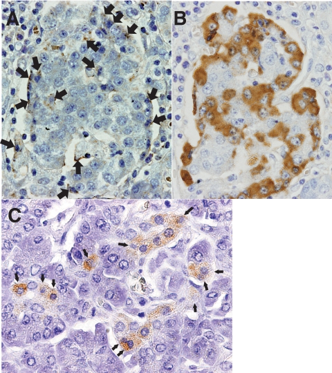
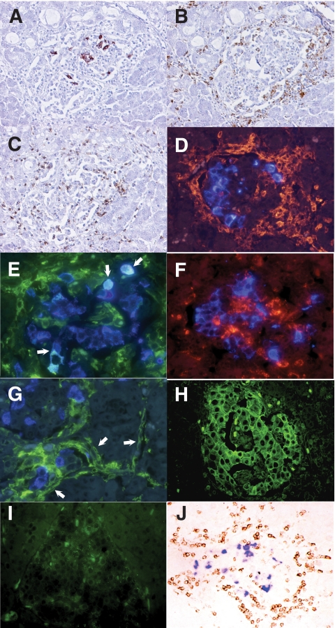
Research design and methods: Subjects comprised three autopsied patients who died from diabetic ketoacidosis within 2-5 days after onset of fulminant type 1 diabetes. We examined islet cell status, including the presence of enterovirus and chemokine/cytokine/major histocompatibility complex (MHC) expressions in the pancreata using immunohistochemical analyses and RT-PCR.
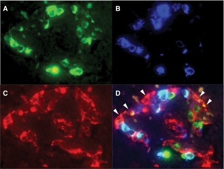
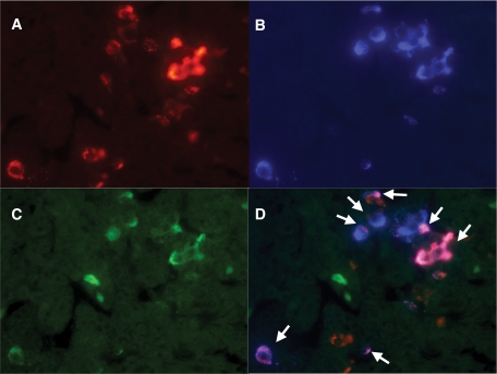
Results: Immunohistochemical analysis revealed the presence of enterovirus-capsid protein in all three affected pancreata. Extensive infiltration of CXCR3 receptor-bearing T-cells and macrophages into islets was observed. Dendritic cells were stained in and around the islets. Specifically, interferon-gamma and CXC chemokine ligand 10 (CXCL10) were strongly coexpressed in all subtypes of islet cells, including beta-cells and alpha-cells. No CXCL10 was expressed in exocrine pancreas. Serum levels of CXCL10 were increased. Expression of MHC class II and hyperexpression of MHC class I was observed in some islet cells.
Conclusions: These results strongly suggest the presence of a circuit for the destruction of beta-cells in fulminant type 1 diabetes. Enterovirus infection of the pancreas initiates coexpression of interferon-gamma and CXCL10 in beta-cells. CXCL10 secreted from beta-cells activates and attracts autoreactive T-cells and macrophages to the islets via CXCR3. These infiltrating autoreactive T-cells and macrophages release inflammatory cytokines including interferon-gamma in the islets, not only damaging beta-cells but also accelerating CXCL10 generation in residual beta-cells and thus further activating cell-mediated autoimmunity until all beta-cells have been destroyed.
Figures
References
-
- Kobayashi T: Immunology and immunogenetics of type 1 diabetes in Japan. IDF Bulletin 1990;35:34–37
-
- Imagawa A, Hanafusa T, Miyagawa J, Matsuzawa Y: A novel subtype of type 1 diabetes mellitus characterized by a rapid onset and an absence of diabetes-related antibodies: Osaka IDDM Study Group. N Engl J Med 2000;342:301–307 - PubMed
-
- Tanaka S, Kobayashi T, Momotsu T: A novel subtype of type 1 diabetes mellitus. N Engl J Med 2000;342:1835–1837 - PubMed
-
- Imagawa A, Hanafusa T, Uchigata Y, Kanatsuka A, Kawasaki E, Kobayashi T, Shimada A, Shimizu I, Toyoda T, Maruyama T, Makino H: Fulminant type 1 diabetes: a nationwide survey in Japan. Diabetes Care 2003;26:2345–2352 - PubMed
-
- Tanaka S, Endo T, Aida K, Shimura H, Yokomori N, Kaneshige M, Furuya F, Amemiya S, Mochizuki M, Nakanishi K, Kobayashi T: Distinct diagnostic criteria of fulminant type 1 diabetes based on serum C-peptide response and HbA1c levels at onset. Diabetes Care 2004;27:1936–1941 - PubMed
Publication types
MeSH terms
Substances
LinkOut - more resources
Full Text Sources
Other Literature Sources
Medical
Research Materials
