. 2009 Nov;23(11):2042-51.
doi: 10.1038/leu.2009.153. Epub 2009 Jul 30.

Novel epigenetic mechanisms that control pluripotency and quiescence of adult bone marrow-derived Oct4(+) very small embryonic-like stem cells

Affiliations

Novel epigenetic mechanisms that control pluripotency and quiescence of adult bone marrow-derived Oct4(+) very small embryonic-like stem cells

D M Shin et al. Leukemia. 2009 Nov.

Abstract

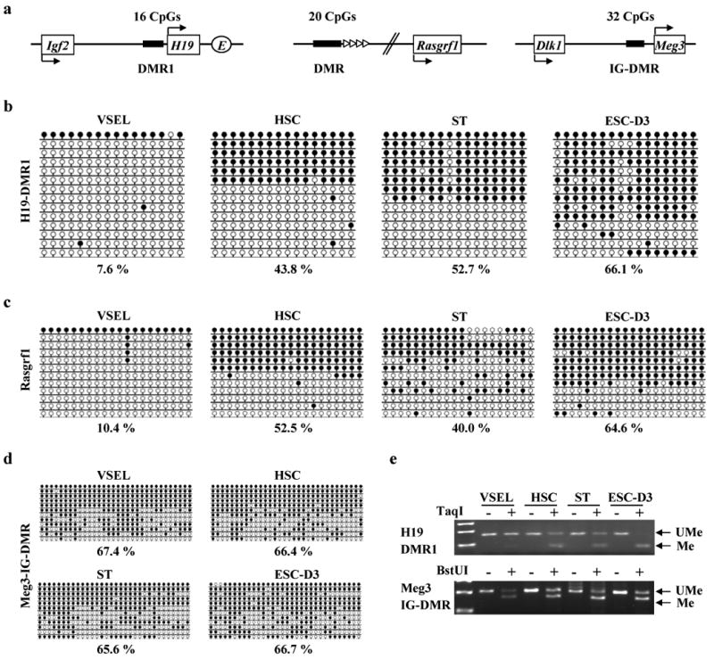
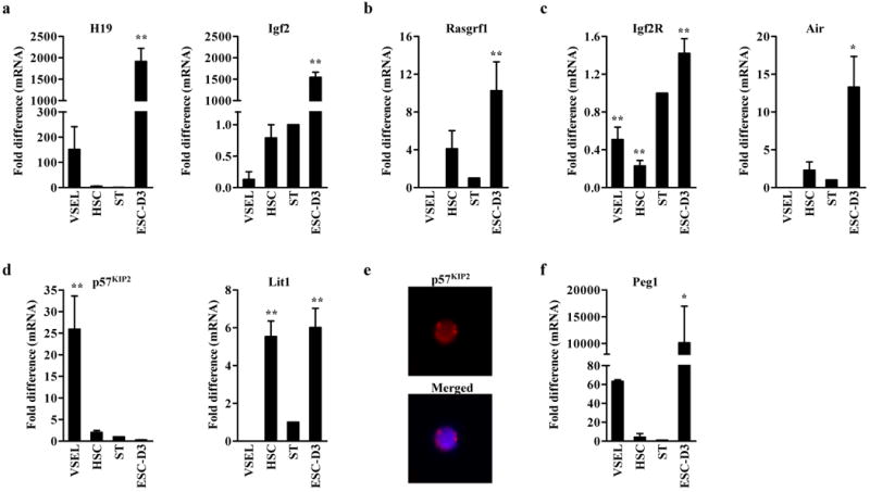
Recently, we identified in adult tissues a population of Oct4(+)SSEA-1(+)Sca-1(+)lin(-)CD45(-) very small embryonic-like stem cells (VSELs). First, to address recent controversies on Oct4 expression in cells isolated from adult organs, we show here evidence that Oct4 promoter in bone marrow (BM)-derived VSELs has an open chromatin structure and is actively transcribed. Next, to explain VSELs quiescence and lack of teratoma formation, we demonstrate a unique DNA methylation pattern at some developmentally crucial imprinted genes, showing hypomethylation/erasure of imprints in paternally methylated and hypermethylation of imprints in maternally methylated ones. These epigenetic characteristics leading to upregulation in VSELs of H19 and p57(KIP2) (also known as Cdkn1c) and repression of Igf2 and Rasgrf1 explain VSEL's quiescent status. Interestingly, this unique pattern in imprinted gene methylation is reverted in cocultures with a C2C12 supportive cell-line when VSELs are induced to form VSEL-derived spheres (VSEL-DSs) enriched for stem cells able to differentiate into all three germ layers. Therefore, we suggest that the proliferative/developmental potential of Oct4(+) VSELs is epigenetically regulated by expression of Oct4 and some imprinted genes, and postulate that restoring the proper methylation pattern of imprinted genes will be a crucial step for using these cells in regenerative medicine.


Conflict of interest statement

Conflicts of Interest Statement: The authors declare that they have no competing financial interests.

Figures

Figure 1
Figure 1. The Oct4 promoter in VSELs is transcriptionally active
(a) The strategy of isolation of VSELs (Lin-Sca-1+CD45-) and HSCs (Lin-Sca-1+CD45+) from murine BM. Note that the lymphocyte gate (R1) was extended to the left to include small sized SCs. (b) Expression of Oct4 mRNA in indicated cells. β-Actin was used as an endogenous housekeeping gene. Control reaction were performed without RTase (-) (c) Immunostaining of Oct4 (Red) and SSEA-1 (Green) protein in VSELs. Note that Oct4 was localized in the nucleus (DAPI blue staining). (d) CpG sites (open-circles) in Oct4 promoter and the target sites of primers (Oct4-S1 and Oct4-S2) employed for the ChIP assay. (e) Bisulfite-sequencing results of DNA methylation of the Oct4 promoter in indicated cells. Methylated and unmethylated CpG sites are shown in filled and open circles, respectively. The numbers under the bisulfite-sequencing profiles indicate the percentage of methylated CpG sites. Regular (f) and quantitative (g) ChIP analyses for the Oct4 promoter in indicated cells to evaluate its association with H3Ac and H3K9me2 histones. In regular ChIP analysis, the amplification of the β-Actin promoter was performed as a control reaction of the endogenous housekeeping gene. Rabbit IgG antibody was used as negative control of immunoprecipitation. In the quantitative ChIP assay, the enrichment of each histone modification was calculated as the ratio of the value from the bound fraction (B) to that from the unbound fraction (UB). Fold differences are shown as the mean ± S.D. from at least four independent experiments. *p<0.05, **p<0.01 compared to BMMNC. IgG; Rabbit IgG antibody
Figure 2
Figure 2. Erasure of genomic imprinting for paternally methylated imprinted-genes in VSELs
(a) The schematic diagram of DMRs for Igf2-H19, Rasgrf1, and Meg3 loci. Upper and bottom arrows represent the maternally and paternally initiated transcription of indicated genes respectively. E: Enhancer. Bisulfite-sequencing profiles of DNA methylation of DMRs for Igf2-H19 (b), Rasgrf1 (c), and Meg3 (d) loci. The percentage of methylated CpG sites was shown by employing bisulfite modification and sequencing results. Unlike DMRs of Igf2-H19 and Rasgrf1, there was little difference in DNA methylation for intergenic (IG)-DMR for Meg3 locus. (e) COBRA assay of Igf2-H19 DMR1 by TaqI restriction enzyme (upper panel) and IG-DMR for Meg3 locus by cleaved BstUI restriction enzyme (lower panel). The unmethylated DNA (UMe) was not cleaved in contrast to methylated DNA (Me) because of a sequence change in the site recognized by a restriction enzyme after bisulfite reaction.
Figure 3
Figure 3. VSELs show the hypermethylated status of DMRs in maternally methylated imprinted-genes
(a) The schematic diagram of DMRs for Kcnq1 and Igf2R loci. Note that DMRs for Kcnq1 and Igf2R loci are located in promoter for antisense-transcripts, Lit1 and Air, respectively. Bisulfite-sequencing results of DNA methylation of DMRs for Kcnq1 (b), Igf2R (c), Peg1 (e), and SNRPN (f) loci. The percentage of methylated CpG sites was shown under the bisulfite-sequencing results. (d) COBRA assay of Igf2R DMR2 cleaved by TaqI restriction enzyme (upper panel) and KvDMR by cleaved BstUI restriction enzyme (lower panel). The UMe was not cleaved in contrast to Me because of a sequence change in the site recognized by a restriction enzyme after bisulfite reaction.
Figure 4
Figure 4. The unique genomic imprinting patterns in VSELs affect the expression level of imprinted-genes
RQ-PCR analysis of Igf2-H19 (a) and Rasgrf1 (b), which DMRs were hypomethylated in VSELs, and maternally methylated imprinted-genes, Igf2R (c), p57KIP2 (d), and Peg1 (f). Of note, VSELs express little antisense-transcripts, Air (c) and Lit1 (d) for Igf2R and Kcnq1 loci, respectively. The relative expression level is represented as the fold difference to the value of STs and shown as the mean ± S.D. from at least three independent experiments on different samples of double-sorted VSELs, HSCs, STs, and ESC-D3. *p<0.05, **p<0.01 compared to ST. (e) The immunostaining of p57KIP2 in VSELs. Note that p57KIP2 protein was localized in the nucleus (merged).
Figure 5
Figure 5. VSELs express a high level of Dnmts
(a) RQ-PCR analysis of Dnmt1, 3b, 3a, and related protein Dnmt3L. The relative expression level was represented as the fold difference to the value of STs and shown as the mean ± S.D. from at least three independent experiments performed on double-sorted VSELs, HSCs, STs, and ESC-D3 cells. *p<0.05, **p<0.01 compared to ST. (b) The immunostaining of Dnmt1 and Dnmt3b proteins in VSELs. DAPI staining was performed to visualize the nucleus and the image merged with DAPI (merged) is shown in right panel. Note that both Dnmts were located in the nucleus (merged).
Figure 6
Figure 6. The repressive genomic imprinting is recovered during VSEL-DS formation
(a) Experimental strategy to isolate cells from VSEL-DSs. VSELs freshly isolated from the BM of GFP transgenic mice (GFP-Tg) were used to grow VSEL-DSs. GFP+ cells were sorted from the cultures. (b) Summary for bisulfite-sequencing results (Supplementary Figure 4) of DNA methylation in the imprinted-genes DMRs and the Oct4 promoter in freshly isolated VSELs and VSEL-DSs (5, 7, 11 days). The paternally imprinted DMRs (H19, Rasgrf1) were marked as blue lines and the maternally imprinted DMRs (Igf2R, KvDMR, Peg1) were marked as red lines. The dashed red line indicates the normal methylation status (50%). (c) Proposed model for epigenetic reprogramming of VSELs deposited in adult tissues during development and their potential activation in response to tissue and organ injury. The proliferative potential of VSELs was indicated in blue (means quiescent status) or red (means proliferative status).

References

    1. Evans MJ, Kaufman MH. Establishment in culture of pluripotential cells from mouse embryos. Nature. 1981;292:154–156. - PubMed
    1. Tesar PJ, Chenoweth JG, Brook FA, Davies TJ, Evans EP, Mack DL, et al. New cell lines from mouse epiblast share defining features with human embryonic stem cells. Nature. 2007;448:196–199. - PubMed
    1. Matsui Y, Zsebo K, Hogan BLM. Derivation of pluripotential embryonic stem cells from murine primordial germ cells in culture. Cell. 1992;70:841–847. - PubMed
    1. Takahashi K, Yamanaka S. Induction of Pluripotent Stem Cells from Mouse Embryonic and Adult Fibroblast Cultures by Defined Factors. Cell. 2006;126:663–676. - PubMed
    1. Wernig M, Meissner A, Foreman R, Brambrink T, Ku M, Hochedlinger K, et al. In vitro reprogramming of fibroblasts into a pluripotent ES-cell-like state. Nature. 2007;448:318–324. - PubMed

Publication types

MeSH terms