. 2009 Jul 30;460(7255):599-604.
doi: 10.1038/nature08218.

Pore architecture and ion sites in acid-sensing ion channels and P2X receptors

Affiliations

Pore architecture and ion sites in acid-sensing ion channels and P2X receptors

Eric B Gonzales et al. Nature. .

Abstract

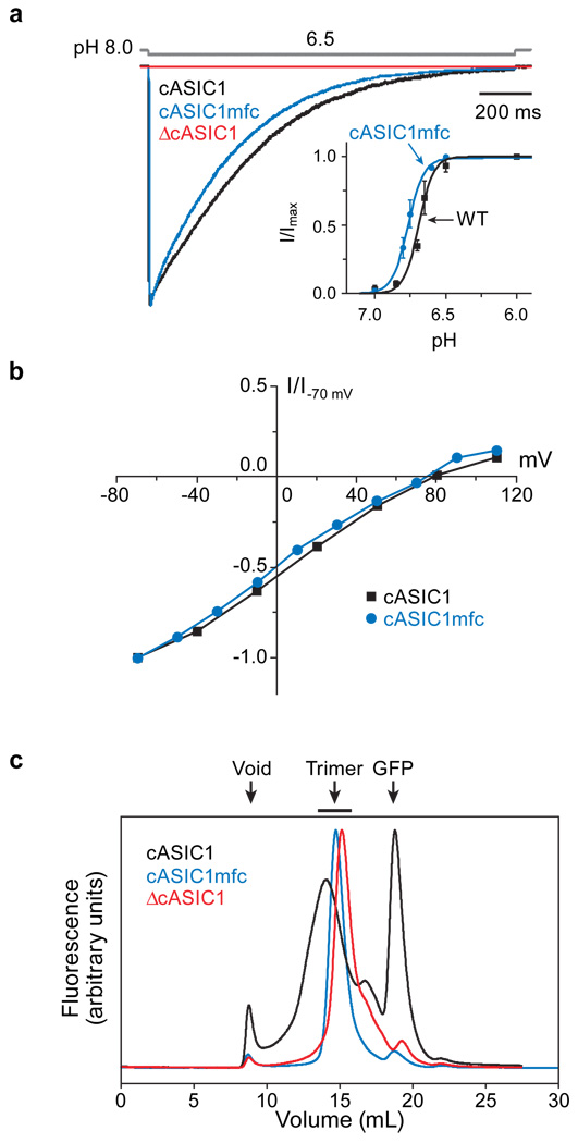
Acid-sensing ion channels are proton-activated, sodium-selective channels composed of three subunits, and are members of the superfamily of epithelial sodium channels, mechanosensitive and FMRF-amide peptide-gated ion channels. These ubiquitous eukaryotic ion channels have essential roles in biological activities as diverse as sodium homeostasis, taste and pain. Despite their crucial roles in biology and their unusual trimeric subunit stoichiometry, there is little knowledge of the structural and chemical principles underlying their ion channel architecture and ion-binding sites. Here we present the structure of a functional acid-sensing ion channel in a desensitized state at 3 A resolution, the location and composition of the approximately 8 A 'thick' desensitization gate, and the trigonal antiprism coordination of caesium ions bound in the extracellular vestibule. Comparison of the acid-sensing ion channel structure with the ATP-gated P2X(4) receptor reveals similarity in pore architecture and aqueous vestibules, suggesting that there are unanticipated yet common structural and mechanistic principles.


Figures

Figure 1
Figure 1. Identification of a minimally functional cASIC1 construct
a, Typical current generated by cASIC1 constructs in outside-out patch recordings (holding potential −60 mV). Channels were activated by stepping from pH 8.0 to 6.5 for the wild-type (black), ΔcASIC1 (red), cASIC1mfc (blue) constructs (open tip junction potential is gray). Typical pH concentration-response relationships are shown in the inset. The wild type channel had pH50 of 6.68 ± 0.01 and n value of 8.35 ± 1.91. The cASIC1mfc had a pH50 and n values of 6.77 ± 0.01 and 7.86 ± 1.22, respectively. Points on the curve are the mean ± s.e.m. of at least four cells. b, Normalized current-voltage relationship. Current observed at low pH and various holding potentials was normalized to the current observed at −70 mV. c, Fluorescence detection size-exclusion chromatography of cASIC1 constructs.
Figure 2
Figure 2. Structure of cASIC1mfc
a, View of the functional, cASIC1mfc trimer. Chloride ions are green spheres. The ‘thumb’, ‘finger’, and ‘wrist’ regions are labelled. b, A vertical slice through a solvent accessible surface representation of the transmembrane domain, the extracellular vestibule and fenestrations. One of the three equivalent fenestrations is indicated by an arrow. Asp433 defines the ‘bottom’ of the extracellular vestibule. c, Key interactions between symmetry related D433 carboxyl and Y425 hydroxyl groups.
Figure 3
Figure 3. Vestibules and possible ion permeation pathways
a, An electrostatic potential surface and cartoon representation of cASIC1mfc sliced along the molecular 3-fold axis of symmetry. The surface is coloured based on electrostatic potential, contoured from −50 kT (red) to +30 kT (blue). White is 0 kT. b, Illustration of the radius of possible pathways along the 3-fold axis, generated using HOLE (red < 1.4 Å < green < 2.3 Å < purple).
Figure 4
Figure 4. Cs+ binding sites
a, Electron density peaks (3.5 σ) from anomalous difference Fourier maps calculated using diffraction data measured from crystals soaked in CsCl. b, Key interactions between Cs+ ions at sites 1 and 2 with the main chain and side chain oxygen atoms of Gly432 and Asp433, respectively. c, Stick representation of cASIC1mfc interaction with Cs+ at site 2. d, Trigonal antiprism coordination of the Cs+ ion by the Gly432 carbonyl and Asp433 carboxyl oxygens. Oxygen atoms (red spheres) form the vertices, while solid lines represent the sides of each of the two staggered triangles of the antiprism.
Figure 5
Figure 5. ASIC and P2X receptors share a common pore architecture
a, Stereoview of cASIC1mfc (purple) and ΔzfP2×4 (gold) crystal structure overlay based on superposition of the respective TM2 domains. b, A structure-based sequence alignment of cASIC1 and zfP2×4 TM2 domains is shown. Corresponding residues in cASIC1mfc and zfP2×4 are highlighted by side chain color. A rat ENaCα sequence is shown for comparison. Views of the cASIC1mfc (c) and ΔzfP2×4 (d) ion channel pores from the extracellular side of the membrane. Selected side chain residues of ΔzfP2×4 are in green. The cASIC1mfc Met438 is modelled as an alanine.

Comment in

References

    1. Waldmann R, Champigny G, Bassilana F, Heurteaux C, Lazdunski M. A proton-gated cation channel involved in acid-sensing. Nature. 1997;386:173–177. - PubMed
    1. Coric T, Zheng D, Gerstein M, Canessa CM. Proton sensitivity of ASIC1 appeared with the rise of fishes by changes of residues in the region that follows TM1 in the ectodomain of the channel. J Physiol. 2005;568:725–735. - PMC - PubMed
    1. Coric T, Passamaneck YJ, Zhang P, Di Gregorio A, Canessa CM. Simple chordates exhibit a proton-independent function of acid-sensing ion channels. FASEB J. 2008;22:1914–1923. - PubMed
    1. Kellenberger S, Schild L. Epithelial sodium channel/degenerin family of ion channels: a variety of functions for a shared structure. Physiol Rev. 2002;82:735–767. - PubMed
    1. Hesselager M, Timmermann DB, Ahring PK. pH Dependency and desensitization kinetics of heterologously expressed combinations of acid-sensing ion channel subunits. J Biol Chem. 2004;279:11006–11015. - PubMed

Publication types

Associated data