Systematic analysis of immune infiltrates in high-grade serous ovarian cancer reveals CD20, FoxP3 and TIA-1 as positive prognostic factors
- PMID: 19641607
- PMCID: PMC2712762
- DOI: 10.1371/journal.pone.0006412
Systematic analysis of immune infiltrates in high-grade serous ovarian cancer reveals CD20, FoxP3 and TIA-1 as positive prognostic factors
Erratum in
- PLoS One. 2013;8(7). doi:10.1371/annotation/976c923b-d991-4a33-b060-cdd8770bdf5d
Abstract
Background: Tumor-infiltrating T cells are associated with survival in epithelial ovarian cancer (EOC), but their functional status is poorly understood, especially relative to the different risk categories and histological subtypes of EOC.
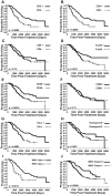
Methodology/principal findings: Tissue microarrays containing high-grade serous, endometrioid, mucinous and clear cell tumors were analyzed immunohistochemically for the presence of lymphocytes, dendritic cells, neutrophils, macrophages, MHC class I and II, and various markers of activation and inflammation. In high-grade serous tumors from optimally debulked patients, positive associations were seen between intraepithelial cells expressing CD3, CD4, CD8, CD45RO, CD25, TIA-1, Granzyme B, FoxP3, CD20, and CD68, as well as expression of MHC class I and II by tumor cells. Disease-specific survival was positively associated with the markers CD8, CD3, FoxP3, TIA-1, CD20, MHC class I and class II. In other histological subtypes, immune infiltrates were less prevalent, and the only markers associated with survival were MHC class II (positive association in endometrioid cases) and myeloperoxidase (negative association in clear cell cases).
Conclusions/significance: Host immune responses to EOC vary widely according to histological subtype and the extent of residual disease. TIA-1, FoxP3 and CD20 emerge as new positive prognostic factors in high-grade serous EOC from optimally debulked patients.
Conflict of interest statement
Figures


References
-
- Ozols RF, Bundy BN, Greer BE, Fowler JM, Clarke-Pearson D, et al. Phase III trial of carboplatin and paclitaxel compared with cisplatin and paclitaxel in patients with optimally resected stage III ovarian cancer: a Gynecologic Oncology Group study. J Clin Oncol. 2003;21:3194–3200. - PubMed
-
- du Bois A, Luck HJ, Meier W, Adams HP, Mobus V, et al. A randomized clinical trial of cisplatin/paclitaxel versus carboplatin/paclitaxel as first-line treatment of ovarian cancer. J Natl Cancer Inst. 2003;95:1320–1329. - PubMed
-
- Holschneider CH, Berek JS. Ovarian cancer: epidemiology, biology, and prognostic factors. Semin Surg Oncol. 2000;19:3–10. - PubMed
-
- Ozols RF. Management of advanced ovarian cancer consensus summary. Advanced Ovarian Cancer Consensus Faculty. Semin Oncol. 2000;27:47–49. - PubMed
-
- Zhang L, Conejo-Garcia JR, Katsaros D, Gimotty PA, Massobrio M, et al. Intratumoral T cells, recurrence, and survival in epithelial ovarian cancer. N Engl J Med. 2003;348:203–213. - PubMed
Publication types
MeSH terms
Substances
LinkOut - more resources
Full Text Sources
Other Literature Sources
Medical
Research Materials
Miscellaneous

