. 2009 Jul 29;4(7):e6415.
doi: 10.1371/journal.pone.0006415.

Chemosensory cues to conspecific emotional stress activate amygdala in humans

Affiliations

Chemosensory cues to conspecific emotional stress activate amygdala in humans

Lilianne R Mujica-Parodi et al. PLoS One. .

Abstract

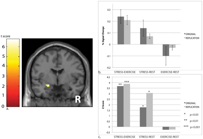
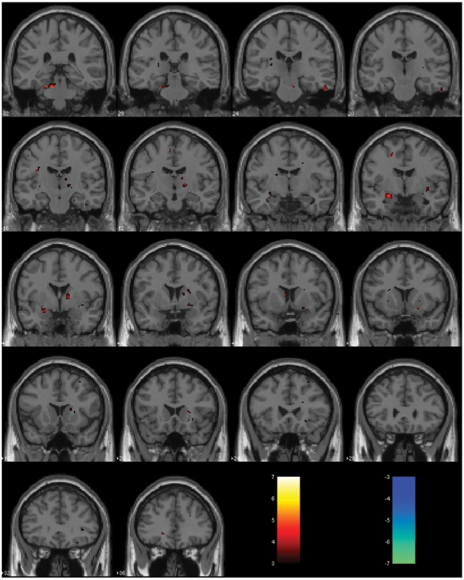
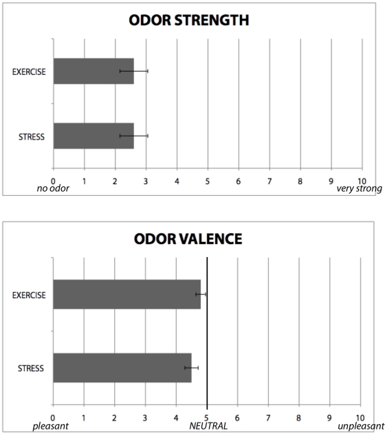
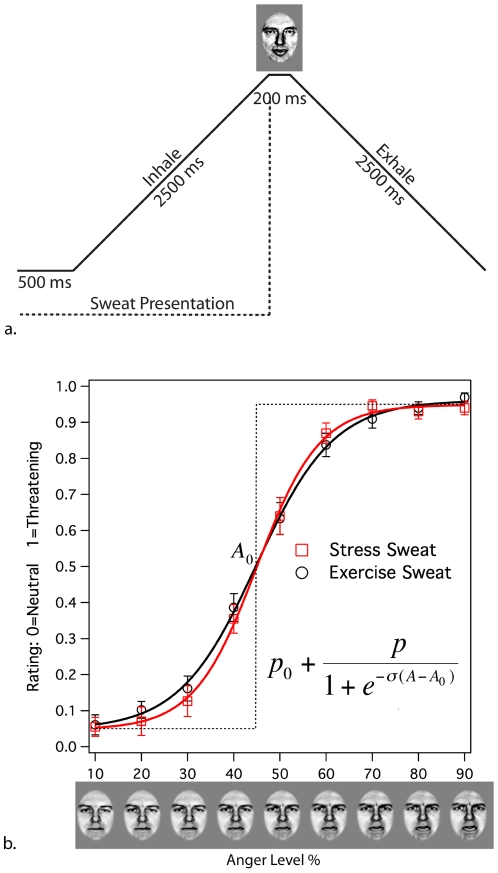
Alarm substances are airborne chemical signals, released by an individual into the environment, which communicate emotional stress between conspecifics. Here we tested whether humans, like other mammals, are able to detect emotional stress in others by chemosensory cues. Sweat samples collected from individuals undergoing an acute emotional stressor, with exercise as a control, were pooled and presented to a separate group of participants (blind to condition) during four experiments. In an fMRI experiment and its replication, we showed that scanned participants showed amygdala activation in response to samples obtained from donors undergoing an emotional, but not physical, stressor. An odor-discrimination experiment suggested the effect was primarily due to emotional, and not odor, differences between the two stimuli. A fourth experiment investigated behavioral effects, demonstrating that stress samples sharpened emotion-perception of ambiguous facial stimuli. Together, our findings suggest human chemosensory signaling of emotional stress, with neurobiological and behavioral effects.


Conflict of interest statement

Competing Interests: The authors have declared that no competing interests exist.

Figures

Figure 1
Figure 1. During the fMRI scans, participants' breathing was synchronized via a continuously expanding and contracting circle (a), which cued inhalation and exhalation, respectively.
Stress and exercise sweat were presented in a randomized block design, with each 20s block comprised of four inhalations-exhalations (b), timed to a five-second cycle.
Figure 2
Figure 2. Breathing stress-derived sweat modulates the amygdala, the primary brain region associated with emotional processing.
The unmasked activation map (a) reflects the STRESS−EXERCISE contrast, and was produced using height threshold t = 3.7, p<0.001 (uncorrected) and extent threshold k = 5 voxels. The MNI coordinates of the maximally activated voxel, located in the left amygdala, are [x = −27, y = −6, z = −12] (t = 5.21/Z = 3.88; p (small-volume-corrected) = 0.008). Inspection of the mean response to STRESS-REST and EXERCISE-REST contrasts (b) initially appeared to suggest mean deactivation in response to EXERCISE sweat. However, once we factored in the variance (c), it became clear that the effect for the STRESS-EXERICISE contrast was predominantly due to activation in response to the STRESS condition, rather than to deactivation in response to the EXERCISE condition, as only the former showed statistically significant changes from baseline.
Figure 3
Figure 3. Full-brain activation maps for replication fMRI study, showing activation levels (STRESS>EXERCISE) in warm colors and de-activations (EXERCISE>STRESS) in cool colors, showed that differences between the two conditions were most pronounced in the amygdala, with no significant de-activations.
These images were produced at p<0.005, with extent threshold = 5 voxels. Table 1 provides a list of all significantly activated clusters corresponding to this whole-brain random-effects analysis.
Figure 4
Figure 4. On Likert Scales, participants rated both conditions as mild and neutral; there were no significant differences between their ratings between conditions.
A separate forced-choice discrimination experiment additionally indicated that participants were unable to distinguish between the two odors. Together, these suggest that the amygdala activation seen in response to the STRESS, but not EXERCISE, sweat was due to engagement of emotional processing rather than perception of distinct odors.
Figure 5
Figure 5. Psychometric curves generated by a forced-choice assessment of ambiguous threat show sharpened discrimination between threat and non-threat while breathing stress-derived sweat.
For each participant, data for each condition (STRESS, EXERCISE) were fitted with the sigmoid function, where p 0 and p 0p define upper and lower asymptotes, A0 is the inflection point, and σ defines slope. Significant differences between conditions were seen for slope, with individuals under the STRESS condition more closely approximating ideal perceptual discrimination, shown by the dotted line.
Figure 6
Figure 6. Gas chromatography mass spectroscopy analyses of exercise sweat samples were used to validate that our collection and aqueous extraction methods were capable of sampling over hydrophobic (steroid) components in human apocrine sweat.
Total ion count gas chromatography trace of aqueous human sweat extract shows the presence of cholesterol, which is hydrophobic.
Figure 7
Figure 7. Gas chromatography mass spectroscopy analyses of exercise sweat samples were used to validate that our collection and aqueous extraction methods were capable of sampling over hydrophobic (steroid) components in human apocrine sweat.
Mass spectrum of retention time 19.512 minutes shows the presence of human steroids found in apocrine sweat.
Figure 8
Figure 8. Gas chromatography mass spectroscopy analyses of exercise sweat samples were used to validate that our collection and aqueous extraction methods were capable of sampling over hydrophobic (steroid) components in human apocrine sweat.
Mass spectrum of retention time 20.655 minutes shows the presence of human steroids found in apocrine sweat.

References

    1. Pfeiffer W. Alarm substances. Experientia. 1963;19:113–123. - PubMed
    1. Kiyokawa Y, Shimozuru M, Kikusui T, Takeuchi Y, Mori Y. Alarm pheromone increases defensive and risk assessment behaviors in male rats. Physiol Behav. 2006;87:383–387. - PubMed
    1. Kiyokawa Y, Kikusui T, Takeuchi Y, Mori Y. Mapping the neural circuit activated by alarm pheromone perception by c-Fos immunohistochemistry. Brain Res. 2005;1043:145–154. - PubMed
    1. Inagaki H, Kiyokawa Y, Kikusui T, Takeuchi Y, Mori Y. Enhancement of the acoustic startle reflex by an alarm pheromone in male rats. Physiol Behav. 2008;93:606–611. - PubMed
    1. Wysocki CJ, Preti G. Facts, fallacies, fears, and frustrations with human pheromones. Anat Rec A Discov Mol Cell Evol Biol. 2004;281:1201–1211. - PubMed

Publication types