. 2009 Jul 25:15:1418-28.

TNF-alpha mediated apoptosis plays an important role in the development of early diabetic retinopathy and long-term histopathological alterations

Affiliations

TNF-alpha mediated apoptosis plays an important role in the development of early diabetic retinopathy and long-term histopathological alterations

Antonia M Joussen et al. Mol Vis. .

Abstract

Purpose: The pathophysiology of diabetic retinopathy involves leukocyte adhesion to retinal vasculature, early blood-retinal barrier breakdown, capillary nonperfusion, and endothelial cell death. We investigated the involvement of tumor necrosis factor alpha (TNF-alpha) in diabetes-related histopathological changes in two relevant rodent models.

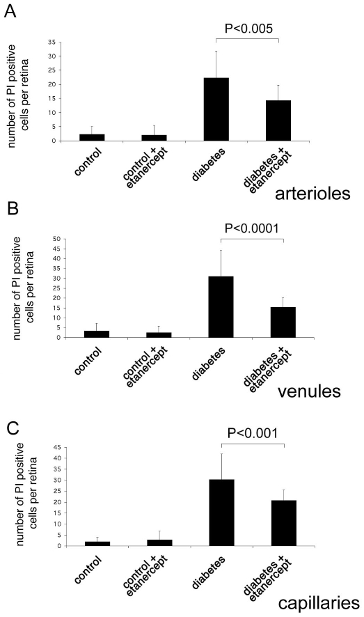
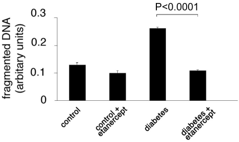
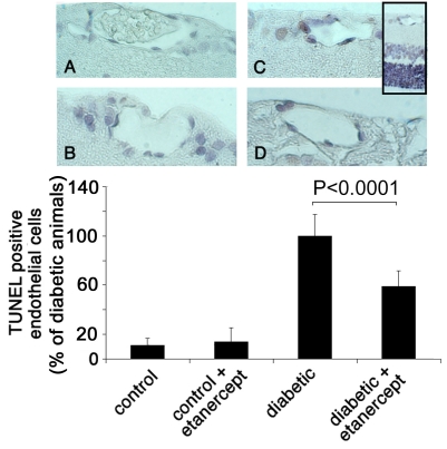
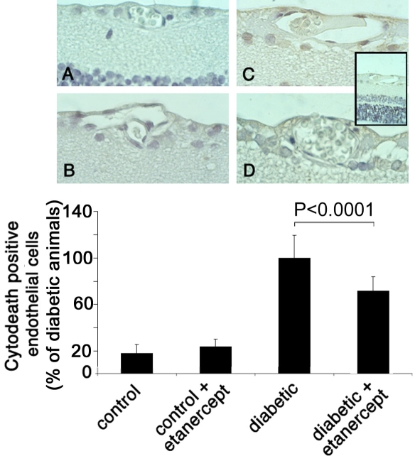
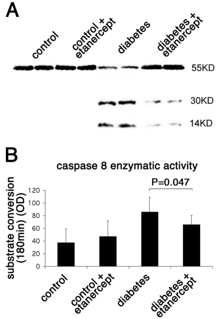
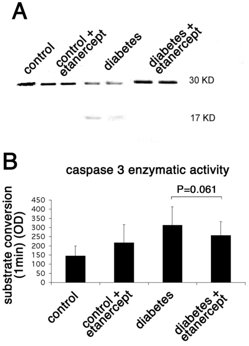
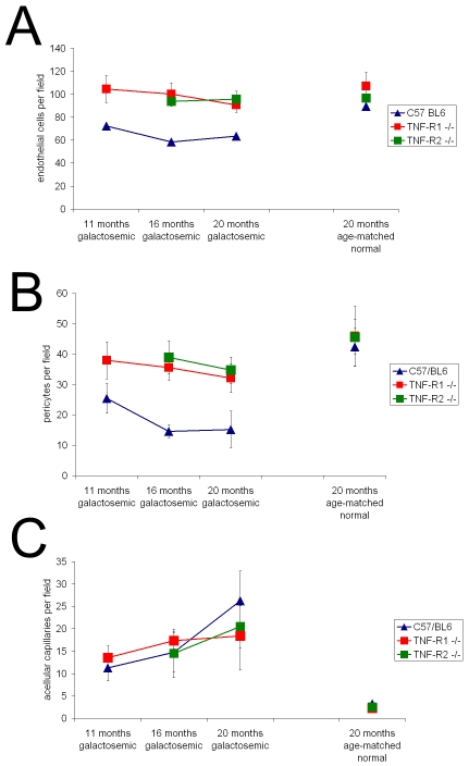
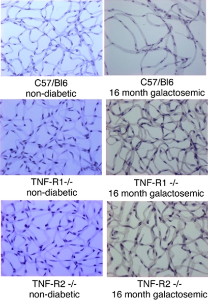
Methods: In short-term studies, Long-Evans rats with streptozotocin-induced diabetes were treated with or without the TNF-alpha inhibitor, etanercept. For long-term studies, tumor necrosis factor receptor I (TNF-RI)-deficient mice and TNF-RII-deficient mice, as well as C57/Bl6 wild-type mice, were fed 30% galactose for up to 20 months. The retinal histopathological alterations of hypergalactosemia were analyzed in trypsin digest preparations. Endothelial cell injury and apoptosis in rat retinas were evaluated by propidium iodide, TUNEL, CytoDeath staining, and DNA fragmentation ELISA. Caspase 3 and 8 activity was evaluated by immunoblotting and quantitative enzymatic activity assay.

Results: Etanercept suppressed caspase activation, retinal cell injury, and apoptosis in short-term diabetic rats. Pericyte and endothelial cell loss were also reduced in long-term hypergalactosemic mice. Long-term studies demonstrated that pericyte loss and endothelial cell loss were reduced in comparison to wild-type diabetic controls.

Conclusions: Our study identifies an important role for TNF-alpha in the pathogenesis of signature diabetic retinopathy pathologies and demonstrates that etanercept can inhibit retinal cell death and long-term complication of diabetes. Taken together, our results suggest that etanercept could prove beneficial in preventing both early and late vascular diabetic complications.


Figures

Figure 1
Figure 1
Quantification of endothelial cell injury and death by propidium iodide staining. Dead and injured retinal endothelial cells were labeled in vivo using propidium iodide. Endothelial cell damage was increased in diabetic animals in capillaries (A), venules (B), and arteries (C). Administration of the TNF-α inhibitor etanercept reduced diabetes-induced endothelial cell injury in all vessel types.
Figure 2
Figure 2
Cellular DNA fragmentation ELISA in the retina. DNA fragmentation and therefore apoptosis was significantly increased in the retinal tissue of diabetic compared to nondiabetic animals. Treatment with etanercept reduced the diabetes-induced increase in DNA fragmentation.
Figure 3
Figure 3
TUNEL staining of retinal sections. Paraffin sections of eyes from nondiabetic rats (A), nondiabetic rats treated with etanercept (B), diabetic rats (C), and diabetic rats treated with etanercept (D) were assessed by TUNEL. Sections were subsequently counterstained with hematoxylin. Insert shows an overview of a diabetic retina with the ganglion cell layer (above), the inner nuclear layer (middle, light blue) and the outer nuclear layer (below, dark blue). Numbers of positive endothelial cells were counted in each of 12 sections and expressed as percent of total endothelial cells. Treatment with etanercept reduced diabetes-induced endothelial cell apoptosis, as detected by TUNEL staining.
Figure 4
Figure 4
M30 CytoDeath staining of retinal sections. Paraffin sections of eyes from non-diabetic rats (A), non-diabetic rats treated with etanercept (B), diabetic rats (C), and diabetic rats treated with etanercept (D) were assessed with M30 staining. Sections were subsequently counterstained with hematoxylin. Insert shows an overview of a diabetic retina with the ganglion cell layer (above), the inner nuclear layer (middle, light blue) and the outer nuclear layer (below, dark blue). Numbers of positive endothelial cells were counted in each of 12 sections and expressed as percent of total endothelial cells. Treatment with etanercept reduced endothelial cell apoptosis and caspase activity as determined by M30 staining.
Figure 5
Figure 5
Caspase 8 enzymatic activity in rat retinas. A: The level of protein expression of caspase 8 in the rat retina was evaluated by western blotting. Diabetes increased the cleaved form of caspase 8 (activated form), whereas treatment with etanercept reduced the diabetes-induced cleavage of caspase 8. B: The enzymatic activity of caspase 8 in the rat retina was evaluated by a colorimetric analysis. Diabetes increased the activity of caspase 8 as measured by the conversion of its specific substrate. Treatment with etanercept reduced the diabetes-induced caspase 8 activation and therefore the conversion of its substrate and correlates with the reduction in the cleaved form of the caspase as measured by the western blot shown.
Figure 6
Figure 6
Caspase 3 enzymatic activity in rat retinas. A: The level of protein expression of caspase 3 in the rat retina was evaluated by western blotting. Diabetes increased the cleaved form of caspase 3 (activated form), whereas treatment with etanercept reduced the diabetes-induced cleavage of caspase 3. B: The enzymatic activity of caspase 3 in the rat retina was evaluated by a colorimetric analysis. Diabetes increased the activity of caspase 3 as measured by the conversion of its specific substrate. Treatment with etanercept reduced the diabetes-induced caspase 3 activation and therefore the conversion of its substrate and correlates with the reduction in the cleaved form of the caspase as measured by the western blot shown.
Figure 7
Figure 7
Retinal histopathological changes were investigated in galactosemic TNF-RI−/− and TNF-RII−/− mice and their respective wild-type controls. Both the number of endothelial cells per field as well as the number of pericytes per field significantly decreased throughout the course of the disease in the wild-type galactosemic animals (A, B). The number of acellular capillaries was significantly increased at 20 months of galactosemia (C).
Figure 8
Figure 8
Representative images of the capillary bed in galacosemic animals and age-matched controls. Histopathological changes were investigated in galactosemic TNF-RI−/− and TNF-RII−/− mice and their respective wild-type controls by trypsin digestion of the retinas. Representative images of the capillary bed of trypsin digested retinas in age-matched controls (left column) and galactosemic animals (right column) are shown.

References

    1. Miyamoto K, Khosrof S, Bursell SE, Rohan R, Murata T, Clermont AC, Aiello LP, Ogura Y, Adamis AP. Prevention of leukostasis and vascular leakage in streptozotocin-induced diabetic retinopathy via intercellular adhesion molecule-1 inhibition. Proc Natl Acad Sci USA. 1999;96:10836–41. - PMC - PubMed
    1. Joussen AM, Murata T, Tsujikawa A, Kirchhof B, Bursell SE, Adamis AP. Leukocyte-mediated endothelial cell injury and death in the diabetic retina. Am J Pathol. 2001;158:147–52. - PMC - PubMed
    1. Engerman RL, Kern TS. Retinopathy in animal models of diabetes. Diabetes Metab Rev. 1995;11:109–20. - PubMed
    1. Kern TS, Engerman RL. Galactose-induced retinal microangiopathy in rats. Invest Ophthalmol Vis Sci. 1995;36:490–6. - PubMed
    1. Kern TS, Engerman RL. A mouse model of diabetic retinopathy. Arch Ophthalmol. 1996;114:986–90. - PubMed

Publication types

MeSH terms