. 2009 Oct;25(7):669-76.
doi: 10.1007/s10554-009-9477-7. Epub 2009 Jul 30.

Right ventricular peak systolic longitudinal strain is a sensitive marker for right ventricular deterioration in adult patients with tetralogy of Fallot

Affiliations

Right ventricular peak systolic longitudinal strain is a sensitive marker for right ventricular deterioration in adult patients with tetralogy of Fallot

Roderick W C Scherptong et al. Int J Cardiovasc Imaging. 2009 Oct.

Abstract

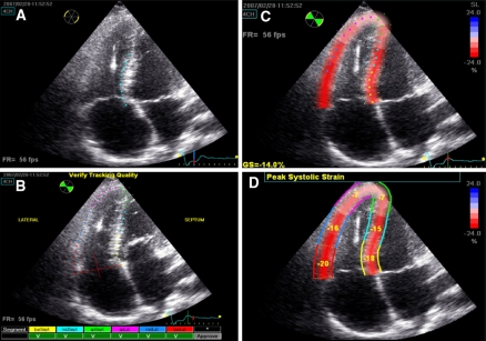
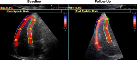
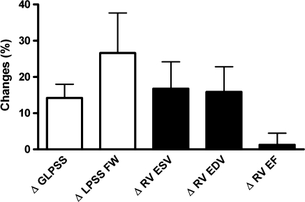
The aim of this study was to evaluate the feasibility of right ventricular (RV) longitudinal peak systolic strain (LPSS) assessment for the follow-up of adult patients with corrected tetralogy of Fallot (TOF). Adult patients (n = 18) with corrected TOF underwent echocardiography and CMR twice with a time interval of 4.2 +/- 1.7 years. RV performance was derived from CMR, and included RV volumes and ejection fraction (EF). LPSS was calculated globally (GLPSS) and in the RV free wall (LPSS FW), with echocardiographic speckle-tracking strain-analysis. Baseline (G)LPSS values were compared between patients and healthy controls; the relation between (G)LPSS and CMR parameters was evaluated and the changes in (G)LPSS and CMR parameters during follow-up were compared. GLPSS and LPSS FW were significantly reduced in patients as compared to controls (-14.9 +/- 0.7% vs. -21.6 +/- 0.9% and -15.5 +/- 0.9% vs. -22.7 +/- 1.5%, P < 0.01). Moderate agreement between LPSS and CMR parameters was observed. RV EF remained unchanged during follow-up, whereas GLPSS and LPSS FW demonstrated a significant reduction. RVEF showed a 1% increase, whereas GLPSS decreased by 14%, and LPSS FW by 27%. RV LPSS is reduced in TOF patients as compared to controls; during follow-up RV EF remained unchanged whereas LPSS decreased suggesting that RV LPSS may be a sensitive marker to detect early deterioration in RV performance.


Figures

Fig. 1
Fig. 1
2D Speckle-tracking Strain Analysis of the Right Ventricle. Right ventricular longitudinal strain analysis in the designated user interface. Panel A demonstrates how first the endocardial border is delineated in the apical 4-chamber view. Based on this contour, the interventricular septum and the right ventricular free wall are tracked automatically. The user optimizes the tracking quality, by adjusting the aforementioned contour and by altering the region of interest (ROI), which corresponds to the width of the tracking (Panel B). Thereafter, the program calculates global strain (GS) based on a weighted average of three septal and three right ventricular free wall segments (Panel C and D)
Fig. 2
Fig. 2
Patient Example of Changes in RV Longitudinal Peak Systolic Strain in Relation to RV Performance on Cardiovascular Magnetic Resonance Imaging. In this patient example, RV end diastolic volume increased gradually from 112 to 120 ml/m2 over a follow-up period of 3 years, but RV ejection fraction remained unchanged (55% at baseline vs. 54% at follow-up). Global RV longitudinal peak systolic strain, however, decreased from −15.5 to −13.6% (red circles), mainly caused by a sharp decrease in longitudinal peak systolic strain of the RV free wall (−17.3% at baseline vs. −12% at 3 years follow-up). These findings indicate that RV performance measured with CMR may exhibit only minimal changes (without change in RV EF), whereas RV LPSS already decreases
Fig. 3
Fig. 3
Changes in Right Ventricular Performance and Right Ventricular Longitudinal Peak Systolic Strain. The white bars represent the overall changes in RV longitudinal peak systolic strain whereas the black bars represent the overall changes in cardiovascular magnetic resonance derived RV volumes and ejection fraction. To compare the relative magnitudes in changes between strain and volumes/ejection fraction, the percentual changes from baseline to follow-up (rather than the absolute changes) are shown. A significant change was noted for RV volumes, global longitudinal peak systolic strain (GLPSS) and longitudinal peak systolic strain in the RV free wall (LPSS FW). The largest changes were observed in LPSS FW

References

    1. Atik FA, Atik E, da Cunha CR et al (2004) Long-term results of correction of tetralogy of Fallot in adulthood. Eur J Cardiothorac Surg 25:250–255 - DOI - PubMed
    1. van Straten A, Vliegen HW, Hazekamp MG et al (2005) Right ventricular function late after total repair of tetralogy of Fallot. Eur Radiol 15:702–707 - DOI - PubMed
    1. Geva T, Sandweiss BM, Gauvreau K et al (2004) Factors associated with impaired clinical status in long-term survivors of tetralogy of Fallot repair evaluated by magnetic resonance imaging. J Am Coll Cardiol 43:1068–1074 - DOI - PubMed
    1. Weidemann F, Eyskens B, Mertens L et al (2002) Quantification of regional right and left ventricular function by ultrasonic strain rate and strain indexes after surgical repair of tetralogy of Fallot. Am J Cardiol 90:133–138 - DOI - PubMed
    1. Gondi S, Dokainish H (2007) Right ventricular tissue Doppler and strain imaging: ready for clinical use? Echocardiography 24:522–532 - DOI - PubMed

Publication types

MeSH terms

LinkOut - more resources