. 2009 Aug 19;20(8):1474-81.
doi: 10.1021/bc800421f. Epub 2009 Jul 31.

Cys-diabody quantum dot conjugates (immunoQdots) for cancer marker detection

Affiliations

Cys-diabody quantum dot conjugates (immunoQdots) for cancer marker detection

Bhaswati Barat et al. Bioconjug Chem. .

Abstract

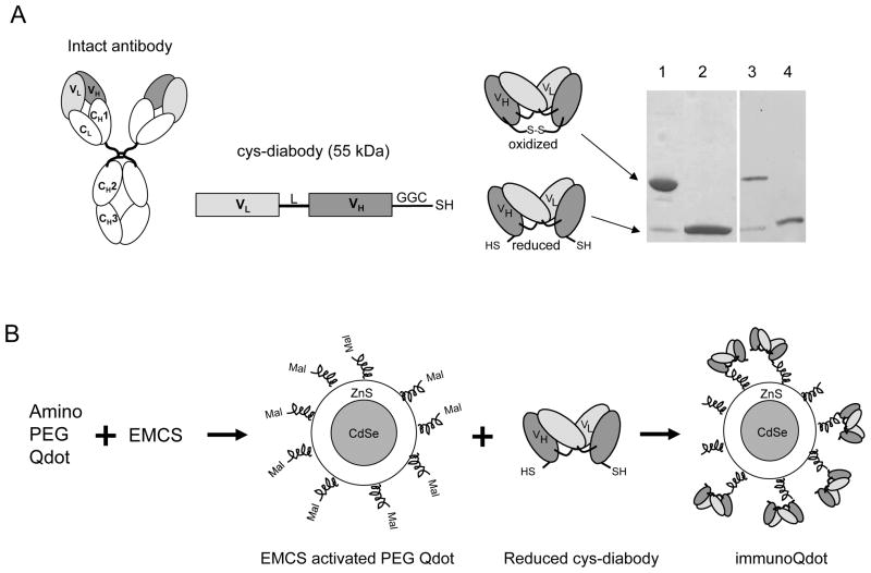
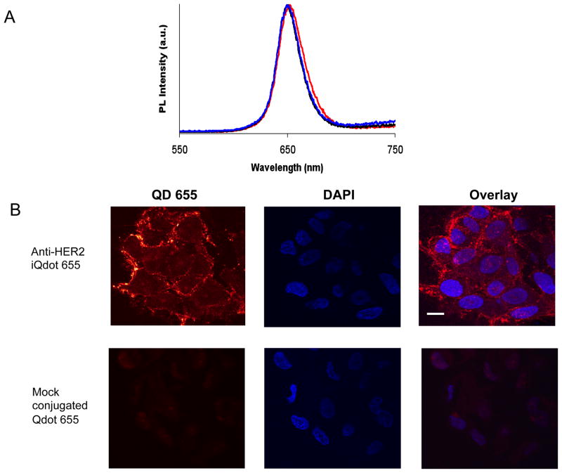
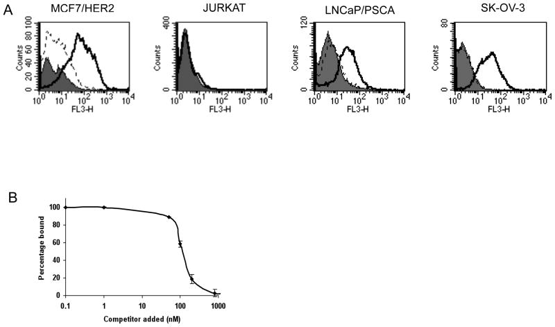
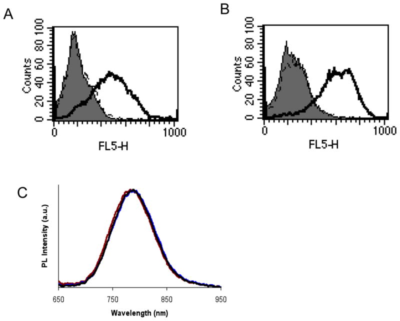
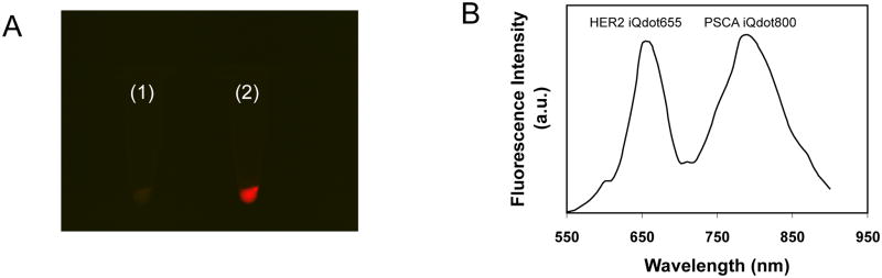
The present work demonstrates the use of small bivalent engineered antibody fragments, cys-diabodies, for biological modification of nanoscale particles such as quantum dots (Qdots) for detection of target antigens. Novel bioconjugated quantum dots known as immunoQdots (iQdots) were developed by thiol-specific oriented coupling of tumor specific cys-diabodies, at a position away from the antigen binding site to amino PEG CdSe/ZnS Qdots. Initially, amino PEG Qdot 655 were coupled with reduced anti-HER2 cys-diabody by amine-sulfhydryl-reactive linker [N-ε-maleimidocaproyloxy] succinimide ester (EMCS) to produce anti-HER2 iQdot 655. Spectral characterization of the conjugate revealed that the spectrum was symmetrical and essentially identical to unconjugated Qdot. Specific receptor binding activity of anti-HER2 iQdot 655 was confirmed by flow cytometry on HER2 positive and negative cells. Immunofluorescence results showed homogeneous surface labeling of the cell membrane with Qdot 655 conjugate. In addition, cys-diabodies specific for HER2, as well as prostate stem cell antigen (PSCA), were conjugated successfully with amino PEG Qdot 800. All of these iQdots retain the photoluminescence properties of the unconjugated Qdot 800 as well as the antigen binding specificity of the cys-diabody as demonstrated by flow cytometry. Simultaneous detection of two tumor antigens on LNCaP/PSCA prostate cancer cells (which express PSCA and HER2) in culture was possible using two iQdots, anti-HER2 iQdot 655 and anti-PSCA iQdot 800. Thus, these iQdots are potentially useful as optical probes for sensitive, multiplexed detection of surface markers on tumor cells. The present thiol-specific conjugation method demonstrates a general approach for site-specific oriented coupling of cys-diabodies to a wide variety of nanoparticles without disturbing the antigen binding site and maintaining small size compared to intact antibody.


Figures

Figure 1
Figure 1
(A) Schematic drawing of an intact antibody showing variable light (VL) and heavy (VH) chain regions and constant (C) regions. Cys-diabody was formed by connecting VL and VH domains with either 5 or 6 amino acid linker (L) and GGC added to the C-termini for cysteine modification. DNA construct and oxidized and reduced form of protein are shown. SDS-PAGE of two cys-diabodies. Lanes 1 and 2, oxidized and reduced form of anti-HER2 cys-diabody; 3 and 4, oxidized and reduced form of anti-PSCA cys-diabody. Non-reduced samples migrate at expected size for covalent dimer (50 kDa) and reduced sample migrate at expected size for monomeric scFv (25 kDa). (B) Schematic illustration of the process of conjugating amino PEG Qdot with cys-diabody. EMCS: [N-e-Maleimidocaproyloxy] succinimide ester. Mal: maleimide group. SDS-PAGE of two Cys-diabodies.
Figure 2
Figure 2
(A) Photoluminescence (emission) spectra of amino PEG Qdot 655 conjugates at excitation wavelength 488 nm. Maximum emission wavelengths are 650.0, 650.5 and 652.5 nm for commercial Qdot 655 (black line), mock conjugated Qdot 655 (blue line) and anti-HER2 iQdot 655 (red line) respectively. All spectra are typically around 30 to 50 nm (full width at half maximum). (B) Confocal microscopy images of MCF7/HER2 cells stained with anti-HER2 iQdot 655 and mock conjugated Qdot 655. Cell nuclei were counterstained with DAPI and shown in blue. Scale bars: 20 μm.
Figure 3
Figure 3
(A) Flow cytometry analysis of cys-diabody conjugated Qdot binding with different tumor cells. Cells were treated with no protein (solid grey), mock conjugated Qdot 655 (dotted black line) and anti-HER2 iQdot 655 (solid black line). FL3 (λem: 670 nm long pass) was the filter used for Qdot 655. (B) Competitive cell binding assay by flow cytometry. An anti-HER2 antibody fragment, minibody (29) was used as competitor. Samples were assayed in triplicate and means ± SEM are shown, normalized to the signal obtained in the absence of competitor.
Figure 4
Figure 4
(A) Flow cytometry analysis of cys-diabody conjugated NIR Qdot binding with MCF7/HER2 Cells. Cells were treated with no protein (solid grey), mock conjugated Qdot 800 (dotted black line) and anti-HER2 iQdot 800 (solid black line). (B) LNCaP/PSCA cells stained with no protein (solid grey), mock conjugated Qdot 800 (dotted black line) and anti-PSCA iQdot 800 (solid black line). FL5 (λem: 740 long pass) was the filter used for Qdot 800. (C) Normalized emission spectra of amino PEG Qdot 800 conjugates. Excitation wavelength was 532 nm. Corresponding emission peaks and associated full-width half-maximum values were 787.9 and 88.95, 785.7 and 89.19, and 789.0 and 89.62 nm for mock conjugated Qdot 655 (black line), anti-HER2 iQdot 800 (red line) and anti-PSCA iQdot 800 (brown line) respectively.
Figure 5
Figure 5
Dual marker Qdot staining of human prostate cancer cells. (A) In vitro fluorescence imaging of LNCaP/PSCA cells stained with (1) mock conjugated Qdot 655 and Qdot 800 and (2) anti-HER2 iQdot 655 and anti-PSCA iQdot 800. The raw fluorescence image acquired using a color CCD camera with a 550 to 900 nm filter is shown. (B) Spectral analysis of dual stained cells (sample 2) following background subtraction.

References

    1. Michalet X, Pinaud FF, Bentolila LA, Tsay JM, Doose S, Li JJ, Sundaresan G, Wu AM, Gambhir SS, Weiss S. Quantum dots for live cells, in vivo imaging, and diagnostics. Science. 2005;307:538–44. - PMC - PubMed
    1. Xing Y, Chaudry Q, Shen C, Kong KY, Zhau HE, Chung LW, Petros JA, O’Regan RM, Yezhelyev MV, Simons JW, Wang MD, Nie S. Bioconjugated quantum dots for multiplexed and quantitative immunohistochemistry. Nat Protoc. 2007;2:1152–65. - PubMed
    1. Bruchez M, Jr, Moronne M, Gin P, Weiss S, Alivisatos AP. Semiconductor nanocrystals as fluorescent biological labels. Science. 1998;281:2013–6. - PubMed
    1. Chan WC, Nie S. Quantum dot bioconjugates for ultrasensitive nonisotopic detection. Science. 1998;281:2016–8. - PubMed
    1. Fountaine TJ, Wincovitch SM, Geho DH, Garfield SH, Pittaluga S. Multispectral imaging of clinically relevant cellular targets in tonsil and lymphoid tissue using semiconductor quantum dots. Mod Pathol. 2006;19:1181–91. - PubMed

Publication types

MeSH terms