. 2009 Oct 15;334(2):335-44.
doi: 10.1016/j.ydbio.2009.07.031. Epub 2009 Jul 28.

Jak-STAT regulation of male germline stem cell establishment during Drosophila embryogenesis

Affiliations

Jak-STAT regulation of male germline stem cell establishment during Drosophila embryogenesis

X Rebecca Sheng et al. Dev Biol. .

Abstract

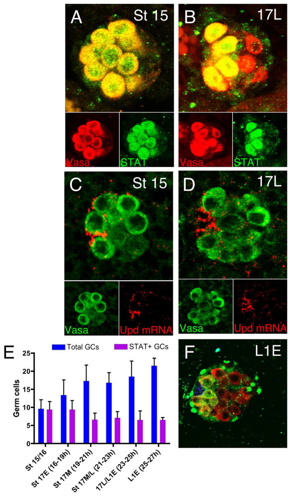
Germline stem cells (GSCs) in Drosophila are descendants of primordial germ cells (PGCs) specified during embryogenesis. The precise timing of GSC establishment in the testis has not been determined, nor is it known whether mechanisms that control GSC maintenance in the adult are involved in GSC establishment. Here, we determine that PGCs in the developing male gonad first become GSCs at the embryo to larval transition. This coincides with formation of the embryonic hub; the critical signaling center that regulates adult GSC behavior within the stem cell microenvironment (niche). We find that the Jak-STAT signaling pathway is activated in a subset of PGCs that associate with the newly-formed embryonic hub. These PGCs express GSC markers and function like GSCs, while PGCs that do not associate with the hub begin to differentiate. In the absence of Jak-STAT activation, PGCs adjacent to the hub fail to exhibit the characteristics of GSCs, while ectopic activation of the Jak-STAT pathway prevents differentiation. These findings show that stem cell formation is closely linked to development of the stem cell niche, and suggest that Jak-STAT signaling is required for initial establishment of the GSC population in developing testes.


Figures

Figure 1
Figure 1. Jak-STAT signaling becomes restricted to anterior germ cells in late stage 17 embryos
(A,B) Wild-type embryos immunostained for STAT92E (green) and Vasa (red) after gonad coalescence. Anterior of the gonads is to the left. Insets show individual channels. (A) Gonad from male stage 15 embryo showing STAT92E accumulation in the nucleus and cytoplasm of all PGCs. (B) Late stage 17 male gonad with STAT92E accumulation restricted to the anterior-most germ cells. (C,D) Fluorescent in situ hybridization performed on wild-type embryos with an anti-sense probe to upd mRNA (red), along with immunostaining for Vasa (green). (C) Stage 15 male gonad with upd expression slightly enriched at the anterior. (D) Late stage 17 male wth upd detected robustly in a tight cluster of SGPs at the gonad anterior (E) Graph showing the total number of germ cells (blue bars) and the number of germ cells with STAT92E immunoreactivity (purple bars) in the gonad collected at various times. (F) Early L1 gonad immunostained for phospho-STAT92E and Sox100B (green), DN-Cadherin (blue), and Vasa (red).
Figure 2
Figure 2. E-cadherin and APC2 enrich at the hub/germ cell interface at the embryo to larval transition
(A–C) nos-Gal4/UAS-Ecadherin::GFP embryos immunostained for GFP (red), FasIII (green), and counterstained for DAPI (blue). (A′–C′) GFP channels shown alone, with yellow arrows indicating regions of E-Cadherin enrichment. (A,A′) Early stage 17 male gonad with E-Cadherin present at the cell membrane of germ cells (arrow). (B,B′) late stage 17/early 1st instar and (C,C′) late 1st instar larvae showing E-Cadherin concentrated at the hub/anterior germ cell interface in some germ cells (arrows). (D–E) Stage 16 and late stage 17 embryos immunostained for APC2 (green) and Vasa (red) with hub outlined. (D′,E′) APC2 channel shown alone APC2 is not polarized in stage 16 gonads (D,D′), but becomes slightly enriched at the hub/germ cell interface in late stage 17 embryos (E, E′ arrow). Anterior is left, and scale bars indicate 10μM (A–C) or 15 μM (D–E).
Figure 3
Figure 3. Anterior germ cells orient their divisions with respect to the hub at the end of embryogenesis
w1118 embryos/larva co-immunostained for γ-tubulin (green), FasIII (green), Vasa (red), and PH3 (blue) to mark orientation of dividing germ cells with respect to the A-P axis of the gonad (see methods). Green dashed lines denote the A-P axis with anterior to the left. White lines denote germ cell division orientation. (A–C) Stage 16/early stage 17 embryos have equal numbers of germ cells with divisions oriented perpendicular (blue), intermediate (green), and parallel (purple) to the A-P axis throughout the gonad (see graph; panel C). (A,B) Representative immunostains showing (A) an anteriorly localized germ cell with intermediate division orientation, and (B) two posterior germ cells dividing with perpendicular orientation. (D–F) Late stage 17 and early L1 embryos display germ cells with the majority of divisions oriented perpendicular to the hub (outlined) in anterior regions, while divisions are randomly oriented in all other regions (see graph; panel F). (D,E) Representative immunostains showing (D) two anterior germ cells making perpendicular divisions, or (E) one germ cell in the middle of the gonad with its division plane oriented parallel to the hub. Scale bars indicate 10μM
Figure 4
Figure 4. Spermatogonial differentiation is observed in posterior germ cells shortly after onset of asymmetric germ cell division at the hub
(A–D) w1118 larva immunostained for BAM (green) and Vasa (red). Anterior to the left and presumptive hub outlined. (A) Male stage 17/early 1st instar larval gonad lacking BAM expression. (B,C) Mid and late larval 1st instar testes with BAM expressed in posterior germ cells. (D) 2nd larval instar testes showing BAM expression in germ cells a few cell diameters away from the hub, but absent in posterior germ cells. (E–I) w1118 embryos/larva immunistained for Vasa (green), FasIII (red), 1B1 (red), and counterstained with DAPI (blue). (E) Stage 16 embryo with all germ cells (example outlined) displaying spherical fusomes. (F) Late stage 17 embryo with germ cells showing spherical fusomes, and bar-like fusomes (outlined) connected to a daughter cell. (G) Mid 1st instar larval testes displaying spherical and bar-like fusomes in hub-proximal germ cells, but elongated fusomes (outlined) in posterior germ cells. (H) Mid 2nd instar larval testes with spherical and bar-like fusomes in hub-proximal germ cells, but elongated and branched fusomes (outlined) in posterior germ cells. (I) Graph showing the gradual appearance of differentiated gonial cyst stages over time (see methods) without the depletion of undifferentiated germline cell types. Note: (*) Once a hub is detected via FasIII immunostain, all germ cells adjacent to the hub are denoted GSCs, (″) while germ cells not adjacent to the hub, either single, or attached to a GSC, are denoted as GBs. Scale bar indicates 10μm.
Figure 5
Figure 5. Jak-STAT activation is required for GSC maintenance and promotes germ cell self-renewal
(A–B) Nos::GMA;;STATTS and sib control embryos/larvae immunostained for GFP (Green) to detect germ cells and determine genotype (see methods), FasIII (red), 1B1 (red), and counterstained with DAPI (blue). Samples collected after 24 hrs growth at non-permissive temperature (see methods). (A) 1st instar larval testes from sibling control showing GSCs with spherical spectrosomes adjacent to the hub. (B) STATTS mutant larval 1st instar testes with spermatogonial cells containing branched fusomes near the hub. (C) Quantitative analyses reveal a marked decrease in the average number of GSCs in STATTS mutant testes grown at non-permissive temperature compared to sibling controls (p<0.0001). (D–E) Nos-Gal4/UAS-Upd larvae immunostained for Vasa (red) and either HTS (D, green) or Bam (E, green) to detect spermatogonial differentiation. Individual HTS (D′) and BAM (E′) channels show for clarity. (D,D′) Late 1st instar larval testes lacking presence of branched fusomes. (E,E′) Late 1st instar larval testes lacking BAM expression. Scale bar is indicates 10 μm (D,E).
Figure 6
Figure 6. Schematic of the PGC to GSC transition during Drosophila testes development
(Left gonad) Jak-STAT signaling specifies male GSC identity in all germ cells after gonad coalescence. (Middle gonad) As hub formation occurs toward the end of embryogenesis, restriction of Jak-STAT activity to hub-proximal germ cells establishes GSCs, which are adherent to the hub and divide perpendicularly. Germ cells at the posterior of the gonad have low levels of Jak-STAT activation and initiate differentiation by the mid first instar larval stage (Right gonad). SGPs expressing Upd are dark blue; germ cells with activated STAT92E are yellow; germ cells without activated STAT92E are red; germ cells enriched for Bam are red with green centers, fusomes and spectrosomes are aqua, adhesion molecules are pink, and mitotic spindles are green. Notches in scale represent hours during embryogenesis.

Similar articles

Cited by

References

    1. Aboim AN. Developpement embryonnaire et post-embryonnaire des gonades normales et agametiques de Drosophila melanogaster. Revue Suisse de Zoologie. 1945;52:53–154.
    1. Arbouzova NI, Zeidler MP. JAK/STAT signalling in Drosophila: insights into conserved regulatory and cellular functions. Development. 2006;133:2605–16. - PubMed
    1. Asaoka M, Lin H. Germline stem cells in the Drosophila ovary descend from pole cells in the anterior region of the embryonic gonad. Development. 2004;131:5079–89. - PubMed
    1. Baksa K, Parke T, Dobens LL, Dearolf CR. The Drosophila STAT protein, stat92E, regulates follicle cell differentiation during oogenesis. Developmental Biology. 2002;243:166–175. - PubMed
    1. Bellve AR. Introduction: the male germ cell; origin, migration, proliferation and differentiation. Semin Cell Dev Biol. 1998;9:379–91. - PubMed

Publication types

MeSH terms

LinkOut - more resources