. 2009 Sep;116(9):1762-9.
doi: 10.1016/j.ophtha.2009.04.015. Epub 2009 Jul 29.

Spectral domain optical coherence tomography imaging of geographic atrophy margins

Affiliations

Spectral domain optical coherence tomography imaging of geographic atrophy margins

Srilaxmi Bearelly et al. Ophthalmology. 2009 Sep.

Abstract

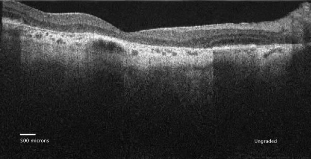
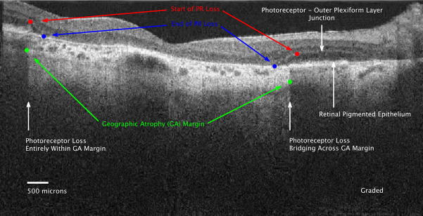
Objective: To test in vivo whether spectral domain optical coherence tomography (SD-OCT) provides adequate resolution for reproducible measurement of photoreceptor (PR) layer at the margins of geographic atrophy (GA), and if it delineates the relationship between PR layer and retinal pigment epithelium at the margins of GA.

Design: Prospective consecutive case series.

Participants: Patients with GA secondary to nonneovascular age-related macular degeneration (AMD) identified during routine follow-up at Duke Eye Center between January 3, 2006, and June 3, 2007, and who consented to participate in this study.

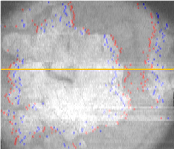
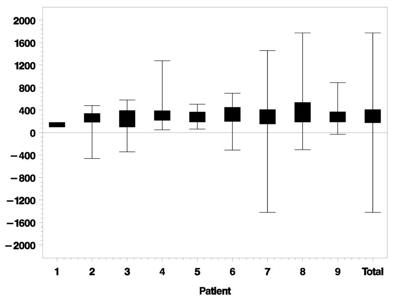
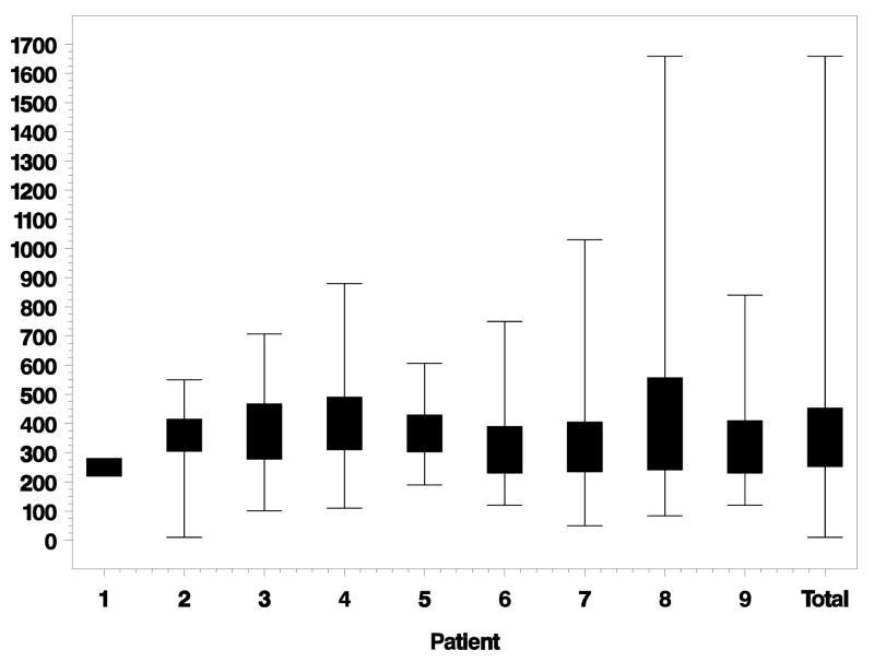
Methods: We used SD-OCT to image eyes. Multiple B-scans from each eye were saved and independently graded by 2 graders and the following locations were marked: (1) site where PR thickness began to decline below its baseline, (2) site where PR layer disappeared, and (3) site of the GA margin. These data were processed to calculate the locations of PR losses relative to GA margins and were categorized as (A) bridging across GA margins, (B) entirely within GA margins, or (C) entirely outside GA margins.

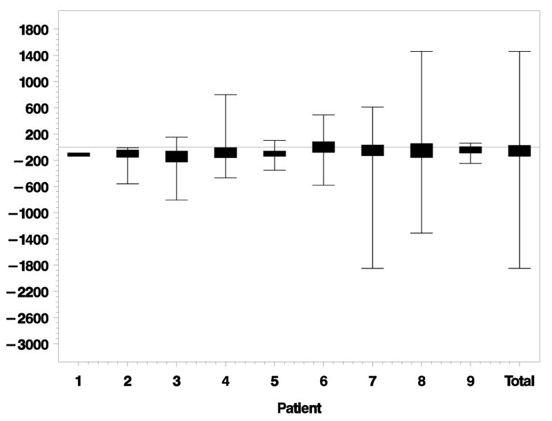
Main outcome measures: Location of PR loss (bridging across GA margins, entirely within GA margins, or entirely outside GA margins) was calculated. Distances from the GA margin were measured for beginning and ending of PR loss. Interobserver agreement was determined for categories of PR loss as well as locations of PR loss relative to the GA margin.

Results: We analyzed 500 unique scans. The PR loss occurred most frequently bridging across the GA margin (65% scans), second most frequently entirely inside the GA margin (29% scans), and least frequently entirely outside the GA margin (6% scans). Loss of PR started an average of 61 microm (standard deviation [SD] +/- 235) outside the GA margin, ended an average of 311+/-273 microm inside the GA margin, and spanned an average of 372+/-179 microm.

Conclusions: Relative to GA margins in non-neovascular AMD with GA, SD-OCT provides adequate resolution for quantifying PR loss. It may also serve as a means of tracking disease progression in future interventional trials.

Financial disclosure(s): Proprietary or commercial disclosure may be found after the references.


Figures

Figure 1
Figure 1
Figure 1a: Ungraded spectral domain optical coherence tomography (SDOCT) image of geographic atrophy (GA) with declining overlying photoreceptor (PR) layer associated with the left and right GA margins. Figure 1b: Graded SD OCT image of GA margin and declining PR layer at the margin. The PR nuclear layer is the hyporeflective (darker) layer between the hyperreflective outer plexiform layer and the retinal pigment epithelium (RPE) (between the white arrows). In the normal eye, the photoreceptor inner segment to outer segment junction is visible as a separate hyperreflective band above the RPE. In this eye, that band is not clearly seen. This is the same image as Figure 1A, but graded features include: 1) Start of PR loss (RED dots), 2) GA margins (GREEN dots), 3) End of PR loss (BLUE dots). The PR layer progressively thins starting at the red dot at each border of the GA until absent at the blue dot. The PR loss is categorized as entirely inside the GA margin on the left, and as bridging across the GA margin on the right. The PR layer is absent between the blue dots, thus the outer plexiform layer is located over Bruch’s membrane. The SDOCT signal is particularly bright in the choroid and extends deeper into the choroid between the green dots where there is no RPE pigment to shadow.
Figure 1
Figure 1
Figure 1a: Ungraded spectral domain optical coherence tomography (SDOCT) image of geographic atrophy (GA) with declining overlying photoreceptor (PR) layer associated with the left and right GA margins. Figure 1b: Graded SD OCT image of GA margin and declining PR layer at the margin. The PR nuclear layer is the hyporeflective (darker) layer between the hyperreflective outer plexiform layer and the retinal pigment epithelium (RPE) (between the white arrows). In the normal eye, the photoreceptor inner segment to outer segment junction is visible as a separate hyperreflective band above the RPE. In this eye, that band is not clearly seen. This is the same image as Figure 1A, but graded features include: 1) Start of PR loss (RED dots), 2) GA margins (GREEN dots), 3) End of PR loss (BLUE dots). The PR layer progressively thins starting at the red dot at each border of the GA until absent at the blue dot. The PR loss is categorized as entirely inside the GA margin on the left, and as bridging across the GA margin on the right. The PR layer is absent between the blue dots, thus the outer plexiform layer is located over Bruch’s membrane. The SDOCT signal is particularly bright in the choroid and extends deeper into the choroid between the green dots where there is no RPE pigment to shadow.
Figure 2
Figure 2
The stack of marked spectral domain optical coherence tomography (SDOCT) scans are summed (averaged) along the z-axis to produce the summed voxel projection (SVP). The multiple sites of markings correspond to the start (RED dots) and end (BLUE dots) of photoreceptor (PR) loss, with the majority of the sites of start of PR loss outside the area of geographic atrophy (GA). The area of absent pigmentation from the retinal pigment epithelium (RPE), i.e. GA, (which corresponds to the area of increased choroidal signal between the green dots in figure 1b) appears white on the SVP. This maps out the area of GA as imaged on SDOCT.
Figure 3
Figure 3
Lateral distance (microns) from beginning of photoreceptor (PR) loss to geographic atrophy (GA) margin for each eye individually. Negative distances are outside the GA margin. Positive distances are inside the GA margin. Eyes ordered by increasing GA size. Total is the combined data of all eyes together.
Figure 4
Figure 4
Lateral distance (microns) from ending of photoreceptor (PR) loss to geographic atrophy (GA) margin for each eye individually. Negative distances are outside the GA margin. Positive distances are inside the GA margin. Eyes ordered by increasing GA size. Total is the combined data of all eyes together.
Figure 5
Figure 5
Lateral distance (microns) from beginning of photoreceptor (PR) loss to ending of PR loss for each eye individually. Eyes ordered by increasing geographic atrophy (GA) size. Total is the combined data of all eyes together.

Comment in

  • Geographic atrophy margins.
    Ergun E, Stur M, Drexler W. Ergun E, et al. Ophthalmology. 2010 May;117(5):1051; author reply 1051. doi: 10.1016/j.ophtha.2010.01.013. Ophthalmology. 2010. PMID: 20438973 No abstract available.

References

    1. Eye Diseases Prevalence Research Group. Prevalence of age-related macular degeneration in the United States. Arch Ophthalmol. 2004;122:564–72. - PubMed
    1. Postel EA, Agarwal A, Caldwell J, et al. Complement factor H increases risk for atrophic age-related macular degeneration. Ophthalmology. 2006;113:1504–7. - PubMed
    1. Sepp T, Khan JC, Thurlby DA, et al. Complement factor H variant Y402H is a major risk determinant for geographic atrophy and choroidal neovascularization in smokers and nonsmokers. Invest Ophthalmol Vis Sci. 2006;47:536–40. - PubMed
    1. Seddon JM, Francis PJ, George S, et al. Association of CFH Y402H and LOC387715 A69S with progression of age-related macular degeneration. JAMA. 2007;297:1793–800. - PubMed
    1. Dunaief JL, Dentchev T, Ying GS, Milam AH. The role of apoptosis in age-related macular degeneration. Arch Ophthalmol. 2002;120:1435–42. - PubMed

Publication types