

Association of white matter hyperintensity measurements on brain MR imaging with cognitive status, medial temporal atrophy, and cardiovascular risk factors
- PMID: 19643919
- PMCID: PMC7051285
- DOI: 10.3174/ajnr.A1693
Association of white matter hyperintensity measurements on brain MR imaging with cognitive status, medial temporal atrophy, and cardiovascular risk factors
Abstract
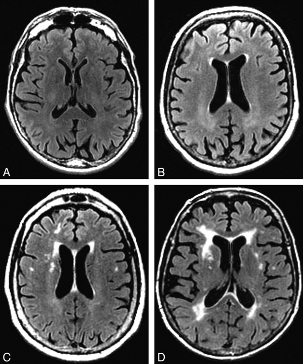
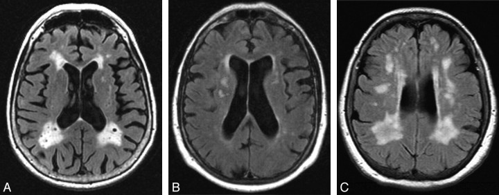
Background and purpose: White matter hyperintensities (WMHs) are frequently characterized as markers of cerebrovascular disease, whereas medial temporal atrophy (MTA) is a recognized marker of Alzheimer disease (AD). Our purpose was to test the reliability of a visual rating system (VRS) in evaluating WMHs and MTA and in distinguishing healthy from cognitively impaired subjects.
Materials and methods: Subjects (n = 192) enrolled in the Florida Alzheimer's Disease Research Center were diagnosed with no cognitive impairment, nonamnestic mild cognitive impairment (na-MCI), amnestic MCI (a-MCI), or probable AD. The severity of WMHs was assessed on T2-weighted fluid-attenuated inversion recovery axial MR images, and the severity of MTA was evaluated on 1.5-mm-thick coronal MR images by using a computer-based visual rating system. Cardiovascular risk factor scores were calculated as the sum of 10 independent cardiovascular risk factors.
Results: WMH and MTA scores were greater in subjects with probable AD, relative to those with no cognitive impairment and na-MCI. MTA scores differentiated subjects with a-MCI from those with no cognitive impairment and na-MCI. The total WMH score was significantly related to MTA (r = 0.39; P < .001) but not to cardiovascular risk factor scores (r = 0.07; P = not significant). The overall correct classification rate of probable AD versus no cognitive impairment by using MTA scores was 81.8%, improving to 86.5% when combined with WMH scores.
Conclusions: Both MTA and WMH scores distinguished subjects with no cognitive impairment and probable AD. Combining MTA and WMH scores improved the correct classification rate, whereas WMH scores were significantly related to MTA scores, but not to cardiovascular risk factor scores. This finding suggests that among subjects with a-MCI and probable AD, WMHs on MR images are primarily associated with neurodegenerative disease.
Figures
References
-
- Kapeller P, Barber R, Vermeulen RJ, et al. Visual rating of age-related white matter changes on magnetic resonance imaging. Stroke 2003;34:441–45 - PubMed
-
- Fazekas F, Kapeller P, Schmidt R, et al. The relation of cerebral magnetic resonance signal hyperintensities to Alzheimer's disease. J Neurolog Sci 1996;142:121–25 - PubMed
-
- Wahlund LO, Barkhof F, Fazekas F, et al. A new scale for age-related white matter changes applicable to MRI and CT. Stroke 2001;32:1318–22 - PubMed
-
- Flier WM, Barkhof F, Scheltens P. Shifting paradigms in dementia. Ann NY Acad Sci 2007;1097:215–24 - PubMed
-
- Rosano C, Alzenstein H, Wu M, et al. Focal atrophy and cerebrovascular disease increase dementia risk among cognitively normal older adults. J Neuroimaging 2007;17:148–55 - PubMed
Publication types
MeSH terms
Grants and funding
LinkOut - more resources
Full Text Sources
Medical