. 2009 Oct;297(4):G781-91.
doi: 10.1152/ajpgi.90605.2008. Epub 2009 Jul 30.

Luminal L-glutamate enhances duodenal mucosal defense mechanisms via multiple glutamate receptors in rats

Affiliations

Luminal L-glutamate enhances duodenal mucosal defense mechanisms via multiple glutamate receptors in rats

Yasutada Akiba et al. Am J Physiol Gastrointest Liver Physiol. 2009 Oct.

Abstract

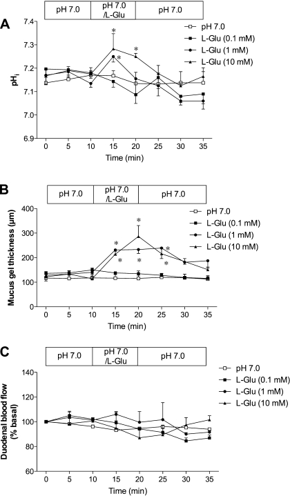
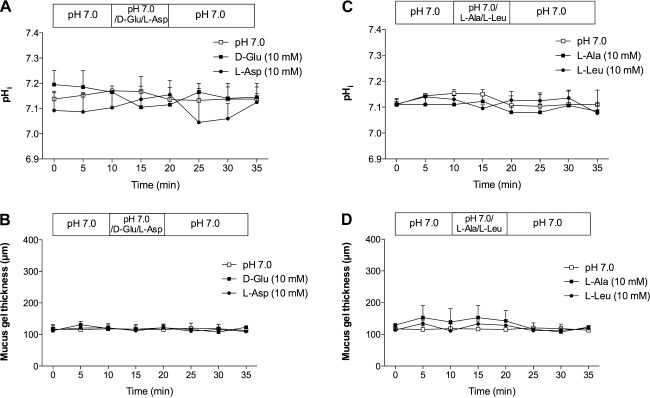
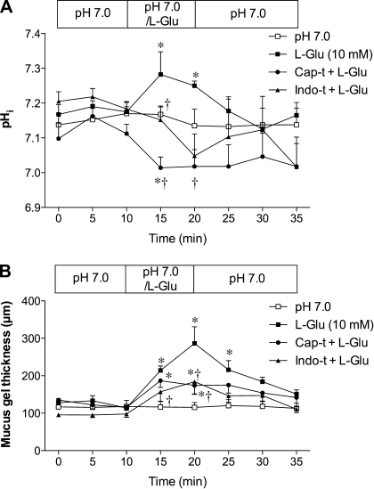
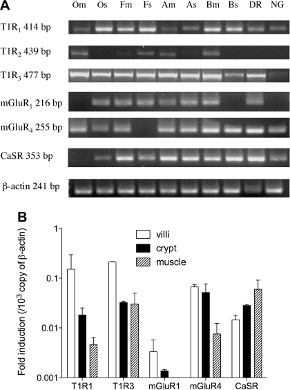
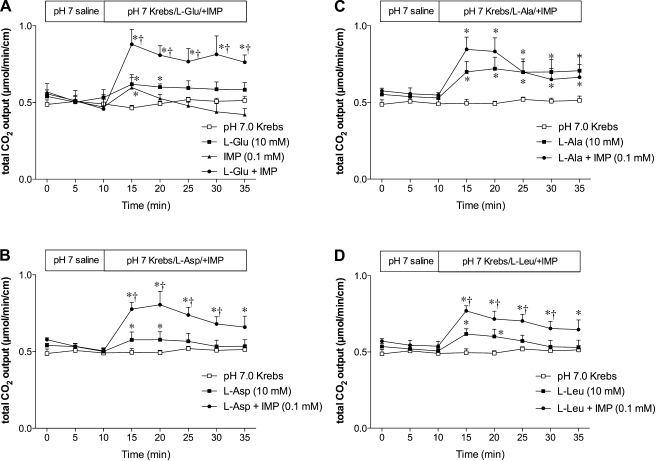
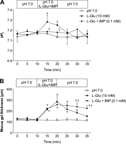
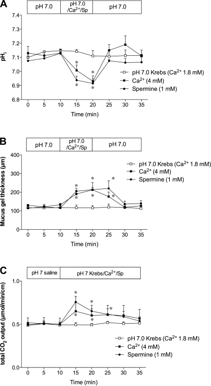
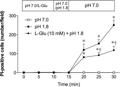
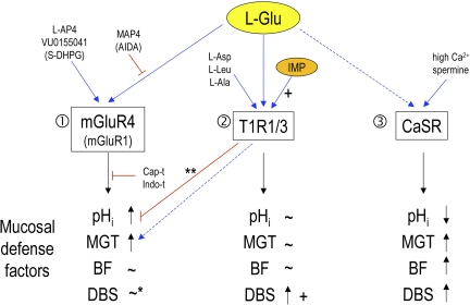
Presence of taste receptor families in the gastrointestinal mucosa suggests a physiological basis for local and early detection of a meal. We hypothesized that luminal L-glutamate, which is the primary nutrient conferring fundamental umami or proteinaceous taste, influences mucosal defense mechanisms in rat duodenum. We perfused the duodenal mucosa of anesthetized rats with L-glutamate (0.1-10 mM). Intracellular pH (pH(i)) of the epithelial cells, blood flow, and mucus gel thickness (MGT) were simultaneously and continuously measured in vivo. Some rats were pretreated with indomethacin or capsaicin. Duodenal bicarbonate secretion (DBS) was measured with flow-through pH and CO(2) electrodes. We tested the effects of agonists or antagonists for metabotropic glutamate receptor (mGluR) 1 or 4 or calcium-sensing receptor (CaSR) on defense factors. Luminal L-glutamate dose dependently increased pH(i) and MGT but had no effect on blood flow in the duodenum. L-glutamate (10 mM)-induced cellular alkalinization and mucus secretion were inhibited by pretreatment with indomethacin or capsaicin. L-glutamate effects on pH(i) and MGT were mimicked by mGluR4 agonists and inhibited by an mGluR4 antagonist. CaSR agonists acidified cells with increased MGT and DBS, unlike L-glutamate. Perfusion of L-glutamate with inosinate (inosine 5'-monophosphate, 0.1 mM) enhanced DBS only in combination, suggesting synergistic activation of the L-glutamate receptor, typical of taste receptor type 1. L-leucine or L-aspartate had similar effects on DBS without any effect on pH(i) and MGT. Preperfusion of L-glutamate prevented acid-induced cellular injury, suggesting that L-glutamate protects the mucosa by enhancing mucosal defenses. Luminal L-glutamate may activate multiple receptors and afferent nerves and locally enhance mucosal defenses to prevent subsequent injury attributable to acid exposure in the duodenum.


Figures

Fig. 1.
Fig. 1.
Effect of luminal l-glutamate (l-Glu) on mucosal defense factors in rat duodenum. Intracellular pH (pHi) of epithelial cells, mucus gel thickness, and blood flow were simultaneously measured in vivo using fluorescence microscopy and flowmetry. Luminal perfusion of l-Glu (0.1–10 mM) dose dependently increased pHi (A) and mucus gel thickness (B) but had no effect on duodenal blood flow (C). Each data point represents mean ± SE (n = 6 rats). *P < 0.05 vs. pH 7.0 Krebs group.
Fig. 2.
Fig. 2.
Effect of luminal amino acids on pHi and mucus gel thickness in rat duodenum. d-glutamate (d-Glu, 10 mM), l-aspartate (l-Asp, 10 mM), l-alanine (l-Ala, 10 mM), or l-leucine (l-Leu, 10 mM) had no effect on pHi (A and C) and mucus gel thickness (B and D). Each data point represents mean ± SE (n = 6 rats).
Fig. 3.
Fig. 3.
Effect of afferent denervation or cyclooxygenase inhibition on l-Glu-induced intracellular alkalinization and mucus secretion in rat duodenum. Rats were pretreated with capsaicin (Cap-t, 125 mg/kg sc) or indomethacin (Indo-t 5 mg/kg, sc). A: pHi. Cap-t and Indo-t inhibited l-Glu-induced intracellular alkalinization, while l-Glu acidified the cells in Cap-t rats. Each data point represents mean ± SE (n = 6 rats). *P < 0.05 vs. pH 7.0 Krebs group, †P < 0.05 vs. l-Glu group. B: mucus gel thickness. Cap-t and Indo-t reduced l-Glu-induced mucus secretion. Each data point represents mean ± SE (n = 6 rats). *P < 0.05 vs. pH 7.0 Krebs group, †P < 0.05 vs. l-Glu group.
Fig. 4.
Fig. 4.
Expression of l-Glu receptors in rats. A: RT-PCR analysis for taste receptor 1 subtypes (T1R1, 2, 3), metabotropic glutamate receptor subtypes (mGluR1, 4), and calcium-sensing receptor (CaSR) in the mucosa (m) and muscle layer (s) of the esophagus (Ö), fundic stomach (F), gastric antrum (A), proximal duodenum (B), dorsal root ganglia (DR) and nodose ganglia (NG) in rats. 1 of 3 experiments is represented. B: real-time PCR for T1Rs, mGluRs, and CaSR in the villi, crypt, and muscle layer of the proximal duodenum. Relative expression of each receptor for β-actin was calculated from threshold cycle (Ct) values. Each data point presents mean ± SE (n = 3 rats).
Fig. 5.
Fig. 5.
Effect of amino acid and inosine monophosphate (IMP) on duodenal HCO3 secretion in rats. Duodenal HCO3 secretion is expressed as total CO2 output. Perfusion of l-Glu (10 mM) or IMP (0.1 mM) had a little effect on HCO3 secretion, while l-Glu coperfused with IMP synergistically augmented HCO3 secretion (A). Similar synergism was observed with perfusion of l-Asp (10 mM) (B) or l-Leu (10 mM) (D), whereas l-Ala (10 mM) increased HCO3 secretion without the synergy with IMP (C). Each data point represents mean ± SE (n = 6 rats). *P < 0.05 vs. pH 7.0 Krebs group, †P < 0.05 vs. the corresponding amino acid group.
Fig. 6.
Fig. 6.
Effect of l-Glu and IMP on pHi and mucus gel thickness in rats. A: coperfusion of IMP (0.1 mM) with l-Glu (10 mM) inhibited l-Glu-induced intracellular alkalinization. Each data point represents mean ± SE (n = 6 rats). *P < 0.05 vs. pH 7.0 Krebs group, †P < 0.05 vs. l-Glu group. B: IMP had no enhancing effect on l-Glu-induced mucus secretion. Each data point represents mean ± SE (n = 6 rats). *P < 0.05 vs. pH 7.0 Krebs group, †P < 0.05 vs. l-Glu group.
Fig. 7.
Fig. 7.
Effect of mGluR agonists or antagonists on duodenal mucosal defense factors. A and B: effects of (S)-3,5-dihydroxyphenylglycine (S-DHPG) (0.1 mM), l-2-amino-4-phosphonobutyric acid (l-AP4) (0.1 mM), or VU0155041 (10 μM) on pHi of epithelial cells (A) and mucus gel thickness (B) were examined in rat duodenum. Each data point represents mean ± SE (n = 6 rats). *P < 0.05 vs. pH 7.0 Krebs group. C: duodenal loop was perfused, and HCO3 secretion was measured with pH and CO2 electrodes. HCO3 secretion is expressed as total CO2 output. Each data point represents mean ± SE (n = 6 rats). D and E: effects of 1-amino-2,3-dihydro-1H-indene-1,5-dicarboxylic acid (AIDA) (0.1 mM), or (S)-2-amino-2-methyl-4-phosphonobutyric acid (MAP4) (0.1 mM) on l-Glu (10 mM)-induced pHi (D) and mucus gel thickness (E) increases were examined. Each data point represents mean ± SE (n = 6 rats). *P < 0.05 vs. pH 7.0 Krebs group, †P < 0.05 vs. l-Glu group.
Fig. 8.
Fig. 8.
Effect of CaSR agonists on duodenal mucosal defense factors. A and B: effects of perfusion of high Ca2+ solution (4 mM) or spermine (Sp) (1 mM) on pHi of epithelial cells (A) and mucus gel thickness (B) were examined in rat duodenum. pH 7.0 Krebs solution containing 1.8 mM Ca2+ was perfused as control. Each data point represents mean ± SE (n = 6 rats). *P < 0.05 vs. pH 7.0 Krebs group. C: effect of Ca2+ (4 mM) or spermine (1 mM) on duodenal HCO3 secretion measured via duodenal loop perfusion was examined. Each data point represents mean ± SE (n = 6 rats). *P < 0.05 vs. pH 7.0 Krebs group.
Fig. 9.
Fig. 9.
Effect of l-Glu on acid-induced epithelial injury in rat duodenum. Epithelial injury was assessed in vivo in situ by propidium iodide (PI) staining. Pretreatment with luminal l-Glu (10 mM) reduced pH 1.8 acid-induced increase of PI-positive cell number. Each data point represents mean ± SE (n = 6 rats). *P < 0.05 vs. pH 7.0 Krebs group, †P < 0.05 vs. l-Glu group.
Fig. 10.
Fig. 10.
Summary diagram: effects of luminal l-Glu on mucosal defense factors in rat duodenum. 1) l-Glu increases pHi and mucus gel thickness (MGT) mainly via mGluR4 without changes of blood flow (BF), mimicked by l-AP4 and VU0155041, inhibited by MAP4, capsaicin pretreatment (Cap-t), or indomethacin (Indo-t). l-Glu-induced effects may involve mGluR1, partially mimicked by S-DHPG and reduced by AIDA. *Note that l-Glu does not likely increase duodenal bicarbonate secretion (DBS) via mGluRs. 2) l-Glu increases DBS, enhanced by IMP, probably via the T1R1/3 heterodimer, mimicked by l-Asp, l-Leu, or l-Ala. l-Glu-induced changes in pHi and MGT are unlikely to be mediated by T1R1/3. **Note that IMP inhibits l-Glu-induced pHi increase and sustained l-Glu-induced MGT increase. 3) CaSR activation by high Ca2+ solution or spermine decreases pHi and increases MGT, BF, and DBS, not mirroring l-Glu-induced effects. Blue arrows, stimulation; red lines, inhibition; +, positive allosteric effect by IMP; dashed line, possible involvement; parenthesis, partial involvement.

References

    1. Adibi SA, Mercer DW. Protein digestion in human intestine as reflected in luminal, mucosal, and plasma amino acid concentrations after meals. J Clin Invest 52: 1586–1594, 1973 - PMC - PubMed
    1. Akiba Y, Furukawa O, Guth PH, Engel E, Nastaskin I, Kaunitz JD. Acute adaptive cellular base uptake in rat duodenal epithelium. Am J Physiol Gastrointest Liver Physiol 280: G1083–G1092, 2001 - PubMed
    1. Akiba Y, Furukawa O, Guth PH, Engel E, Nastaskin I, Kaunitz JD. Sensory pathways and cyclooxygenase regulate mucus gel thickness in rat duodenum. Am J Physiol Gastrointest Liver Physiol 280: G470–G474, 2001 - PubMed
    1. Akiba Y, Furukawa O, Guth PH, Engel E, Nastaskin I, Sassani P, Dukkipatis R, Pushkin A, Kurtz I, Kaunitz JD. Cellular bicarbonate protects rat duodenal mucosa from acid-induced injury. J Clin Invest 108: 1807–1816, 2001 - PMC - PubMed
    1. Akiba Y, Ghayouri S, Takeuchi T, Mizumori M, Guth PH, Engel E, Swenson ER, Kaunitz JD. Carbonic anhydrases and mucosal vanilloid receptors help mediate the hyperemic response to luminal CO2 in rat duodenum. Gastroenterology 131: 142–152, 2006 - PubMed

Publication types

MeSH terms

Substances

LinkOut - more resources