

Review
doi: 10.1007/s12105-009-0121-6.
Epub 2009 Jun 10.
Chondromyxoid fibroma of sphenoid sinus with unusual calcifications: case report with literature review
Affiliations
- PMID: 19644549
- PMCID: PMC2715466
- DOI: 10.1007/s12105-009-0121-6
Item in Clipboard
Review
Chondromyxoid fibroma of sphenoid sinus with unusual calcifications: case report with literature review
Head Neck Pathol.
2009 Jun.
Abstract
Chondromyxoid fibroma (CMF) is a rare benign primary tumor which usually affects the metaphyses of the long bone of the lower extremities in childhood and young adults. Rarely, CMF occurs in the skull base and parasinuses, which may be difficult to distinguish from chondrosarcoma or chordoma and other tumors in the head. It is composed of chondroid, myxoid, and fibrous tissue growth in a lobular pattern, infrequently with calcifications. We report one case of CMF involving the sphenoid sinus mimicking a chondrosarcoma. The tumor mass showed calcifications on images and histology.
Keywords: Calcification; Chondromyxoid fibroma; Sphenoid sinus.
Figures
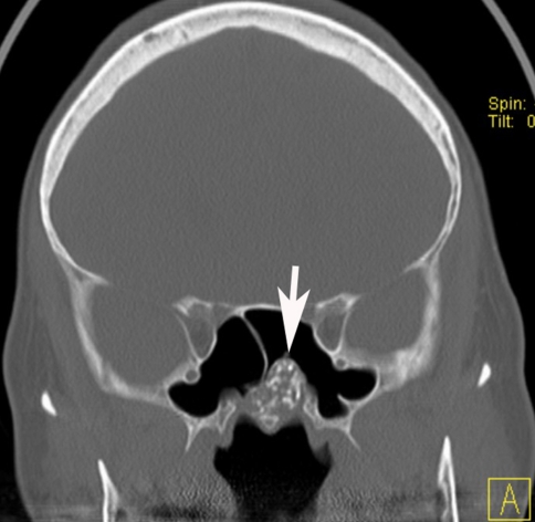
A non-contrast CT scan of the paranasal sinuses showing a heterogeneous mass with fine calcifications, involving the left sphenoid sinus. The mass partially eroded the floor of the sphenoid sinus and protruded into the left nasal cavity (arrow)
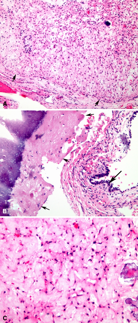
Low magnification of tumor mass shows a well circumscribed cartilaginous neoplasm, with lobular border and peripheral hypercellularity (arrowheads) (a). Section shows the tumor (arrowheads) is adjacent to the sphenoid cavity lined by sinonasal mucosa (arrow), with calcification on the left side (b). High magnification shows tumor is composed of chondromyxoid matrix with stellate fibromyoblastic cells and calcifications (c)
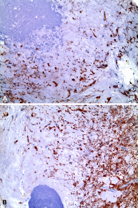
Immunostains show tumor cells were positive for vimentin (a) and smooth muscle actin (b)
Similar articles
-
Sinonasal Chondromyxoid Fibroma: Case Report and Literature Review.Cureus. 2019 Oct 5;11(10):e5841. doi: 10.7759/cureus.5841. Cureus. 2019. PMID: 31754576 Free PMC article. Review.
-
Sphenoid sinus chondromyxoid fibroma mimicking a mucocele.Am J Otolaryngol. 2006 Nov-Dec;27(6):406-8. doi: 10.1016/j.amjoto.2006.01.004. Am J Otolaryngol. 2006. PMID: 17084225
-
Myxoid chondrosarcoma of the sphenoid sinus and chondromyxoid fibroma of the iliac bone: cytomorphologic findings of two distinct and uncommon myxoid lesions.Diagn Cytopathol. 2000 Jun;22(6):383-9. doi: 10.1002/(sici)1097-0339(200006)22:6<383::aid-dc11>3.0.co;2-h. Diagn Cytopathol. 2000. PMID: 10820534
-
Sinonasal chondromyxoid fibroma.Ann Diagn Pathol. 2009 Feb;13(1):41-6. doi: 10.1016/j.anndiagpath.2007.05.006. Epub 2007 Oct 24. Ann Diagn Pathol. 2009. PMID: 19118781
-
[Chondromyxoid fibroma of the nasal septum].Otolaryngol Pol. 2010 Mar-Apr;64(2):88-92. doi: 10.1016/S0030-6657(10)70041-X. Otolaryngol Pol. 2010. PMID: 20568536 Review. Polish.
Cited by
-
Chondromyxoid Fibroma of the Skull Base and Calvarium: Surgical Management and Literature Review.J Neurol Surg Rep. 2016 Mar;77(1):e023-34. doi: 10.1055/s-0035-1570033. Epub 2016 Jan 4. J Neurol Surg Rep. 2016. PMID: 26929898 Free PMC article.
-
Chondromyxoid fibroma of the temporal bone: A rare case report.Medicine (Baltimore). 2020 Mar;99(11):e19487. doi: 10.1097/MD.0000000000019487. Medicine (Baltimore). 2020. PMID: 32176085 Free PMC article. Review.
-
Sinonasal Chondromyxoid Fibroma: Case Report and Literature Review.Cureus. 2019 Oct 5;11(10):e5841. doi: 10.7759/cureus.5841. Cureus. 2019. PMID: 31754576 Free PMC article. Review.
-
[Chondromyxoid fibroma of the mandible: a case report].Hua Xi Kou Qiang Yi Xue Za Zhi. 2016 Dec 1;34(6):654-656. doi: 10.7518/hxkq.2016.06.020. Hua Xi Kou Qiang Yi Xue Za Zhi. 2016. PMID: 28318171 Free PMC article. Chinese.
-
Radiological presentation of chondromyxoid fibroma in the sellar region: A CARE-compliant article and literature review.Medicine (Baltimore). 2017 Dec;96(49):e9049. doi: 10.1097/MD.0000000000009049. Medicine (Baltimore). 2017. PMID: 29245307 Free PMC article. Review.
References
-
- Jaffe HL, Lichtenstein L. Chondromyxoid fibroma of bone: a distinctive benign tumor likely to be mistaken especially for chondrosarcoma. Arch Pathol (Chic) 1948;45:541–551. - PubMed
-
- Dahlin DC. Chondromyxoid fibroma in bone tumors: general aspects and data on 6, 221 cases. Spring-field, IL: Charles C. Thomas; 1978. pp. 57–70.
-
- Gherlinzoni F, Rock M, Pucci P. Chondromyxoid fibroma: the experience at the Instituto Ortopedico Rizzoli. J Bone Joint Surg. 1983;65A:198–204. - PubMed
-
- Schajowicz F, Gallardo H, Argentina BA. Chondromyxoid fibroma (fibromyxoid chondroma) of bone: a clinico-pathologic study of 32 cases. J Bone Joint Surg. 1971;53:198–216. - PubMed
Publication types
MeSH terms
LinkOut - more resources
Full Text Sources
