

Aptamer nano-flares for molecular detection in living cells
- PMID: 19645478
- PMCID: PMC3200529
- DOI: 10.1021/nl901517b
Aptamer nano-flares for molecular detection in living cells
Abstract
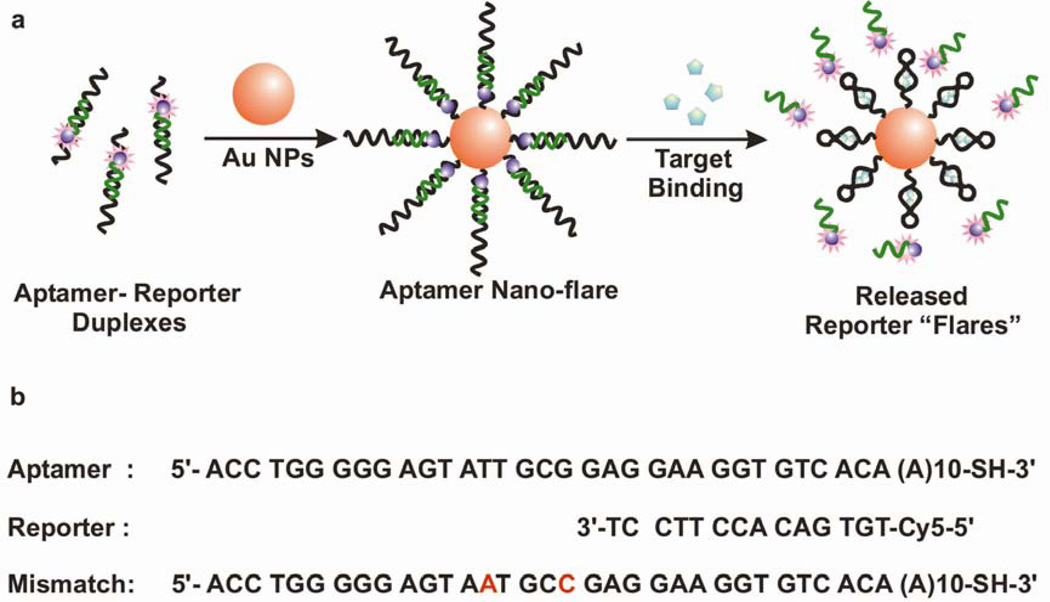
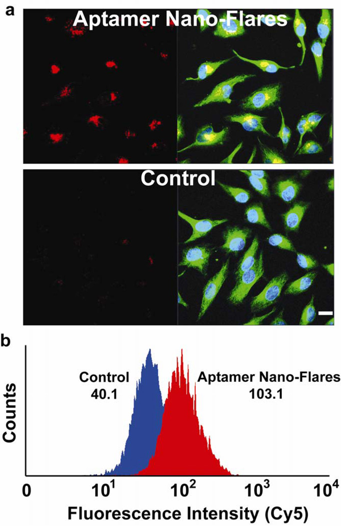
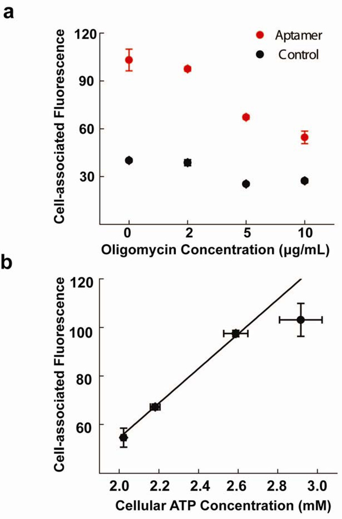
We demonstrate a composite nanomaterial, termed an aptamer nano-flare, that can directly quantify an intracellular analyte in a living cell. Aptamer nano-flares consist of a gold nanoparticle core functionalized with a dense monolayer of nucleic acid aptamers with a high affinity for adenosine triphosphate (ATP). The probes bind selectively to target molecules and release fluorescent reporters which indicate the presence of the analyte. Additionally, these nanoconjugates are readily taken up by cells where their signal intensity can be used to quantify intracellular analyte concentration. These nanoconjugates are a promising approach for the intracellular quantification of other small molecules or proteins, or as agents that use aptamer binding to elicit a biological response in living systems.
Figures

Similar articles
-
Nanoplasmonic detection of adenosine triphosphate by aptamer regulated self-catalytic growth of single gold nanoparticles.Chem Commun (Camb). 2012 Oct 7;48(77):9574-6. doi: 10.1039/c2cc34632j. Chem Commun (Camb). 2012. PMID: 22871726
-
Determination of adenosine triphosphate based on the use of fluorescent terbium(III) organic frameworks and aptamer modified gold nanoparticles.Mikrochim Acta. 2019 Dec 9;187(1):34. doi: 10.1007/s00604-019-4019-z. Mikrochim Acta. 2019. PMID: 31814046
-
Terbium ion-coordinated carbon dots for fluorescent aptasensing of adenosine 5'-triphosphate with unmodified gold nanoparticles.Biosens Bioelectron. 2016 Dec 15;86:978-984. doi: 10.1016/j.bios.2016.07.105. Epub 2016 Jul 30. Biosens Bioelectron. 2016. PMID: 27498324
-
Engineering of switchable aptamer micelle flares for molecular imaging in living cells.ACS Nano. 2013 Jul 23;7(7):5724-31. doi: 10.1021/nn402517v. Epub 2013 Jun 14. ACS Nano. 2013. PMID: 23746078 Free PMC article.
-
Dispersions based on noble metal nanoparticles-DNA conjugates.Adv Colloid Interface Sci. 2011 Apr 14;163(2):123-43. doi: 10.1016/j.cis.2011.02.007. Epub 2011 Feb 19. Adv Colloid Interface Sci. 2011. PMID: 21382609 Review.
Cited by
-
Dynamic DNA assemblies mediated by binding-induced DNA strand displacement.J Am Chem Soc. 2013 Feb 20;135(7):2443-6. doi: 10.1021/ja311990w. Epub 2013 Feb 6. J Am Chem Soc. 2013. PMID: 23360527 Free PMC article.
-
Nucleic-Acid Structures as Intracellular Probes for Live Cells.Adv Mater. 2020 Apr;32(13):e1901743. doi: 10.1002/adma.201901743. Epub 2019 Jul 4. Adv Mater. 2020. PMID: 31271253 Free PMC article. Review.
-
A brief review of cytotoxicity of nanoparticles on mesenchymal stem cells in regenerative medicine.Int J Nanomedicine. 2019 May 24;14:3875-3892. doi: 10.2147/IJN.S205574. eCollection 2019. Int J Nanomedicine. 2019. PMID: 31213807 Free PMC article. Review.
-
Nucleic acid-gold nanoparticle conjugates as mimics of microRNA.Small. 2011 Nov 18;7(22):3158-62. doi: 10.1002/smll.201101018. Epub 2011 Sep 16. Small. 2011. PMID: 21922667 Free PMC article.
-
DNA-Mediated Cellular Delivery of Functional Enzymes.J Am Chem Soc. 2015 Dec 2;137(47):14838-41. doi: 10.1021/jacs.5b09711. Epub 2015 Nov 20. J Am Chem Soc. 2015. PMID: 26587747 Free PMC article.
References
-
- Xiao Y, Patolsky F, Katz E, Hainfeld JF, Willner I. Science. 2003;299(5614):1877–1881. - PubMed
-
- Tkachenko AG, Xie H, Coleman D, Glomm W, Ryan J, Anderson MF, Franzen S, Feldheim DL. J. Am. Chem. Soc. 2003;125(16):4700–4701. - PubMed
-
- Sandhu KK, McIntosh CM, Simard JM, Smith SW, Rotello VM. Bioconj. Chem. 2002;13(1):3–6. - PubMed
-
- Liu J, Lu Y. J. Am. Chem. Soc. 2003;125(22):6642–6643. - PubMed
-
- Wang J, Liu GD, Merkoci A. J. Am. Chem. Soc. 2003;125(11):3214–3215. - PubMed
Publication types
MeSH terms
Substances
Grants and funding
LinkOut - more resources
Full Text Sources
Other Literature Sources
