

Parietal gray matter volume loss is related to spatial processing deficits in long-term abstinent alcoholic men
- PMID: 19645730
- PMCID: PMC2755629
- DOI: 10.1111/j.1530-0277.2009.01019.x
Parietal gray matter volume loss is related to spatial processing deficits in long-term abstinent alcoholic men
Abstract
Background: We previously demonstrated relatively intact cognitive function (with the exception of suggestive evidence for persistent deficits in spatial information processing) in middle-aged long-term abstinent alcoholics (LTAA, abstinent for 6 months or more) compared to age and gender comparable nonalcoholic controls (NAC) (Fein et al., 2006).
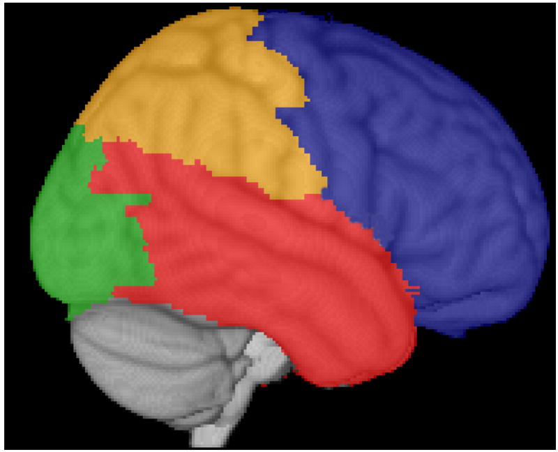
Methods: In the current study, we examine cortical gray matter volumes in the same samples to determine whether gray matter volumes in LTAA are consistent with the cognitive results--i.e., exhibiting gray matter volumes comparable to NAC in most brain regions, except for possible indications of persistent shrinkage in the parietal lobe subserving spatial information processing.
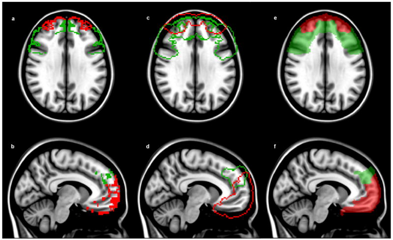
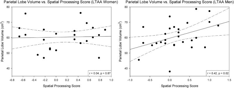
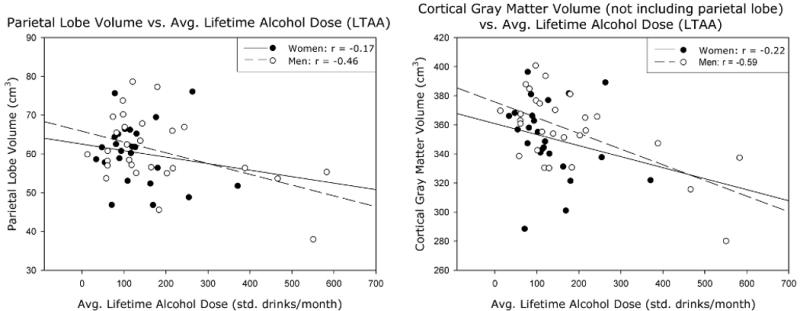
Results: We found gray matter shrinkage in LTAA in the parietal lobe consistent with the spatial processing deficits in this same sample. More compelling, in LTAA, the magnitude of parietal gray matter shrinkage was negatively associated with spatial processing domain performance and positively associated with alcohol dose. Gray matter volume deficits were present in the occipital and other cortical tissue, but poorer visuospatial test performance correlated significantly with smaller volumes in the parietal cortex only.
Conclusions: Taken together, the cognitive and structural imaging data provide compelling evidence that chronic alcohol abuse results in shrinkage of the parietal cortex with associated deficits in spatial information processing.
Figures

Similar articles
-
Association of frontal and posterior cortical gray matter volume with time to alcohol relapse: a prospective study.Am J Psychiatry. 2011 Feb;168(2):183-92. doi: 10.1176/appi.ajp.2010.10020233. Epub 2010 Nov 15. Am J Psychiatry. 2011. PMID: 21078704 Free PMC article.
-
Compromised pontocerebellar and cerebellothalamocortical systems: speculations on their contributions to cognitive and motor impairment in nonamnesic alcoholism.Alcohol Clin Exp Res. 2003 Sep;27(9):1409-19. doi: 10.1097/01.ALC.0000085586.91726.46. Alcohol Clin Exp Res. 2003. PMID: 14506401
-
Cognitive performance in long-term abstinent alcoholic individuals.Alcohol Clin Exp Res. 2006 Sep;30(9):1538-44. doi: 10.1111/j.1530-0277.2006.00185.x. Alcohol Clin Exp Res. 2006. PMID: 16930216 Free PMC article.
-
Smaller gray matter volumes in frontal and parietal cortices of solvent abusers correlate with cognitive deficits.AJNR Am J Neuroradiol. 2009 Nov;30(10):1922-8. doi: 10.3174/ajnr.A1728. Epub 2009 Jul 30. AJNR Am J Neuroradiol. 2009. PMID: 19643925 Free PMC article.
-
White matter (dis)connections and gray matter (dys)functions in visual neglect: gaining insights into the brain networks of spatial awareness.Cortex. 2008 Sep;44(8):983-95. doi: 10.1016/j.cortex.2008.03.006. Epub 2008 May 23. Cortex. 2008. PMID: 18603235 Review.
Cited by
-
Association of frontal and posterior cortical gray matter volume with time to alcohol relapse: a prospective study.Am J Psychiatry. 2011 Feb;168(2):183-92. doi: 10.1176/appi.ajp.2010.10020233. Epub 2010 Nov 15. Am J Psychiatry. 2011. PMID: 21078704 Free PMC article.
-
Mechanisms of postural control in alcoholic men and women: biomechanical analysis of musculoskeletal coordination during quiet standing.Alcohol Clin Exp Res. 2010 Mar 1;34(3):528-37. doi: 10.1111/j.1530-0277.2009.01118.x. Epub 2009 Dec 17. Alcohol Clin Exp Res. 2010. PMID: 20028360 Free PMC article.
-
Impaired fear recognition and attentional set-shifting is associated with brain structural changes in alcoholic patients.Addict Biol. 2014 Nov;19(6):1041-54. doi: 10.1111/adb.12175. Epub 2014 Aug 6. Addict Biol. 2014. PMID: 25123156 Free PMC article.
-
Thinking after Drinking: Impaired Hippocampal-Dependent Cognition in Human Alcoholics and Animal Models of Alcohol Dependence.Front Psychiatry. 2016 Sep 30;7:162. doi: 10.3389/fpsyt.2016.00162. eCollection 2016. Front Psychiatry. 2016. PMID: 27746746 Free PMC article. Review.
-
Towards a Dynamic Exploration of Vision, Cognition and Emotion in Alcohol-Use Disorders.Curr Neuropharmacol. 2019;17(6):492-506. doi: 10.2174/1570159X16666180828100441. Curr Neuropharmacol. 2019. PMID: 30152285 Free PMC article. Review.
References
-
- American Psychiatric Association. DSM-IV-TR: Diagnostic and Statistical Manual of Mental Disorders. Washington, DC: American Psychiatric Publishing; 2000.
-
- Bartsch AJ, Homola G, Biller A, Smith SM, Weijers HG, Wiesbeck GA, Jenkinson M, De Stefano N, Solymosi L, Bendszus M. Manifestations of early brain recovery associated with abstinence from alcoholism. Brain. 2007;130:36–47. - PubMed
-
- Benton AL, Hamsher K. Multilingual aphasia examination. Iowa City: AJA Associates; 1983.
-
- Bucholz KK, Robins LN, Shayka JJ, Przybeck TR, Helzer JE, Goldring E, Klein MH, Greist JH, Erdman HP, Skare SS. Performance of two forms of a computer psychiatric screening interview: version I of the DISSI. Psychiatry Research. 1991;25:117–29. - PubMed
Publication types
MeSH terms
Grants and funding
LinkOut - more resources
Full Text Sources
Medical
