Review
. 2009 May:1164:147-54.
doi: 10.1111/j.1749-6632.2009.03859.x.

Signal processing and distribution in cortical-brainstem pathways for smooth pursuit eye movements

Affiliations
Review

Signal processing and distribution in cortical-brainstem pathways for smooth pursuit eye movements

Michael J Mustari et al. Ann N Y Acad Sci. 2009 May.

Abstract

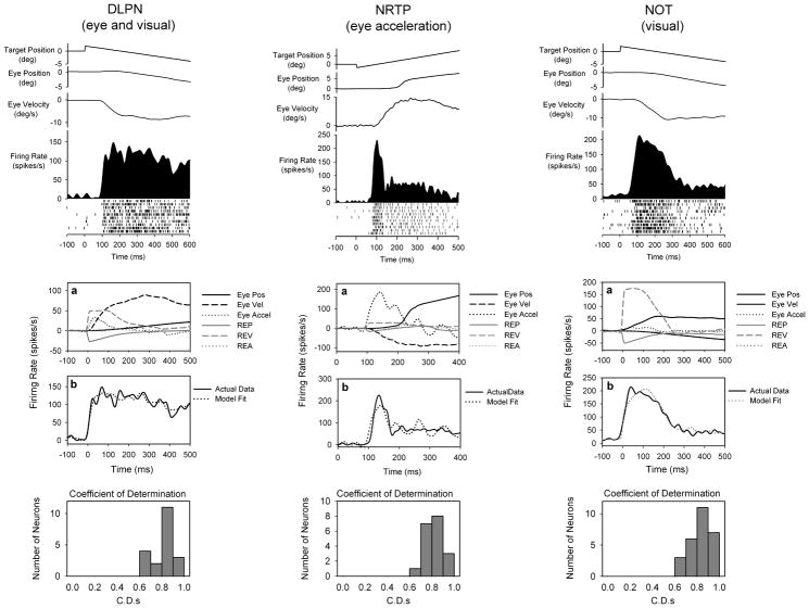
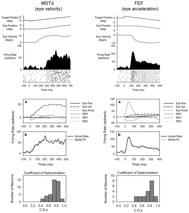
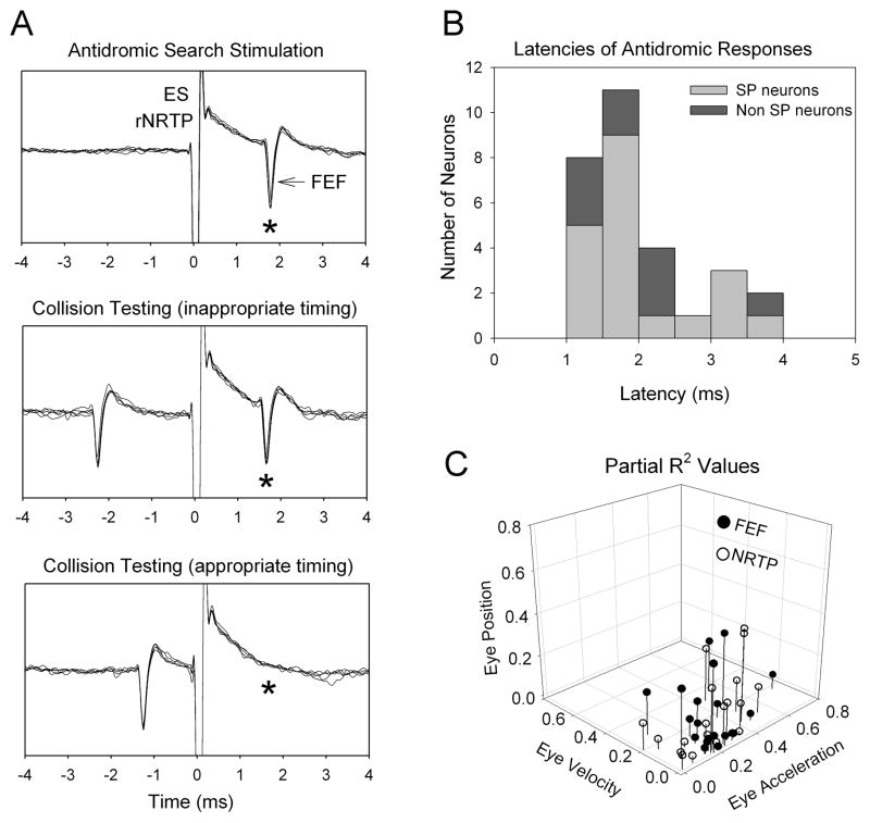
Smooth pursuit (SP) eye movements are used to maintain the image of a moving object relatively stable on the fovea. Even when tracking a single target over a dark background, multiple areas including frontal eye fields (FEF) and middle temporal (MT) and medial superior temporal (MST) cortex contribute to converting visual signals into initial commands for SP. Signals in the cortical pursuit system reach the oculomotor cerebellum through brainstem centers including the dorsolateral pontine nucleus (DLPN), nucleus reticularis tegmenti pontis (NRTP), and pretectal nucleus of the optic tract (NOT). The relative information carried in these parallel pathways remains to be fully defined. We used multiple linear-regression modeling to estimate the relative sensitivities of cortical (MST, FEF), pontine (NRTP, DLPN), and NOT neurons to eye- and retinal-error parameters (position, velocity, and acceleration) during step-ramp SP of macaques (Macaca mulatta). We found that a large proportion of pursuit-related MST and DLPN neurons were most sensitive to eye-velocity or retinal error velocity. In contrast, a large proportion of FEF and rostral NRTP neurons were most sensitive to eye acceleration. Visual neurons in MST, DLPN, and NOT were most sensitive to retinal image velocity.


Figures

Figure 1
Figure 1
Major feed-forward pathways for smooth pursuit (SP). Visual motion is decoded in retinal-genciulo-striate pathways to dorsal stream areas (e.g., MT). Direct retinal and indirect cortical input to the accessory optic system and NOT also provide visual signals for SP. Area MT projects to MST and FEF where different aspects of SP are supported. The cortical SP areas (MT, MST and FEF) project to the pontine nuclei (DLPN and NRTP), which provide parellel channels of information to different cerebellar areas including, flocculus, ventral paraflocculus and vermis. These cerebellar areas enable SP via connections with deep cerebellar nuclei and distal brainstem motor centers. Abbreviations; lateral geniculate nucleus (LGN), dCK (dorsal cap of Kooy inferior olive), vPFloc (ventral paraflocculus), FTN (floccular target neuron), III (oculomotor nucleus), VI (abducens nucleus), MVN (medial vestibular nucleus), NPH (nucleus prepositus hypoglossi), HC, (horizontal semicircular canal).
Figure 2
Figure 2
Examples of smooth pursuit related brainstem neurons. Top panel shows neuronal response of DLPN (left), rNRTP (middle) and NOT (right) neurons during step-ramp SP. Traces show horizontal target and eye position, eye velocity, and neuronal activity (spike density and rasters). (A), Curve fitting with individual traces showing the dynamic values of the components that make up the model. (B), Model fits to neuronal data using 6- or 3-component models. SP neurons in DLPN, rNRTP and NOT are most sensitive to eye velocity, eye acceleration and retinal image velocity, respectively. (C), Histograms of C.D.s for neurons in different regions.
Figure 3
Figure 3
Examples of SP related neurons in MSTd and FEF. MSTd and FEF neurons are most sensitive to eye velocity and eye acceleration, respectively. (conventions as in figure 2).
Figure 4
Figure 4
Characterization of signals in FEF-NRTP pathway. Example of FEF that was antidromically activated following stimulation of rNRTP. Left column (A) shows five successive antidromic trials for each testing condition aligned on the electrical stimulation artifact. Top, search mode reveals antidromic neuron elicited at constant latency. Middle: Antidromic spikes (*) continued to be elicited when inappropriate timing was used between a naturally occurring FEF spike and the stimulus pulse. Bottom panel: when appropriate timing was used between the naturally occurring spike and the stimulus pulse, collision occurs (i.e., no evoked FEF at expected time (*). Antidromic latencies for smooth pursuit and visual neurons in FEF (B). partial-r2 values for FEF and NRTP neurons (C).

References

    1. Leigh RJ, Zee DS. The neurology of eye movements. 4. New York: Oxford Univ. Press; 2006.
    1. Krauzlis RJ. Recasting the smooth pursuit eye movement system. J Neurophysiol. 2004;91:591–603. - PubMed
    1. Voogd J, Barmack NH. Oculomotor Cerebellum. Prog Brain Res. 2006;151:231–268. - PubMed
    1. Newsome WT, Wurtz RH, Dursteler MR, Mikami A. Deficits in visual motion processing following ibotenic acid lesions of the middle temporal visual area of the macaque monkey. J Neurosci. 1985;5:825–840. - PMC - PubMed
    1. Dursteler MR, Wurtz RH, Newsome WT. Directional pursuit deficits following lesions of the foveal representation within the superior temporal sulcus of the macaque monkey. J Neurophysiol. 1987;57:1262–1287. - PubMed

Publication types