. 2009 Jul 31:10:42.
doi: 10.1186/1471-2172-10-42.

In vitro, but not in vivo, reversibility of peritoneal macrophages activation during experimental acute pancreatitis

Affiliations

In vitro, but not in vivo, reversibility of peritoneal macrophages activation during experimental acute pancreatitis

Sabrina Gea-Sorlí et al. BMC Immunol. .

Abstract

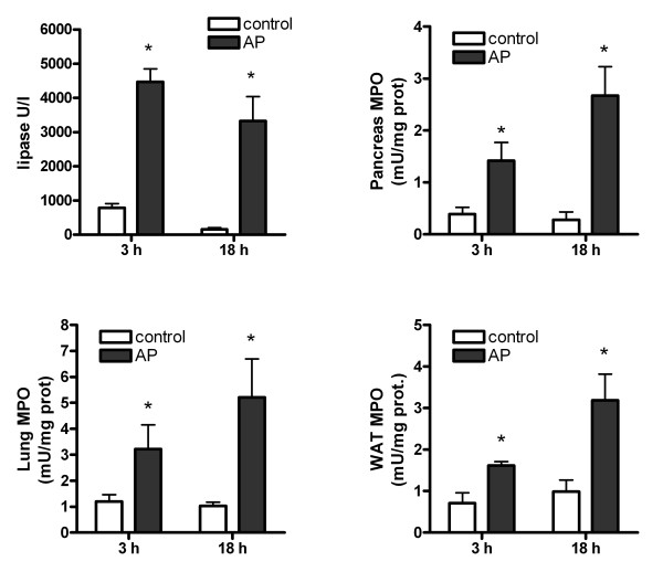
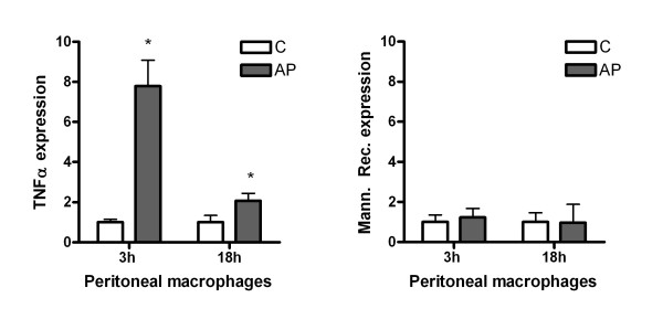
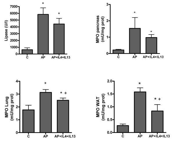
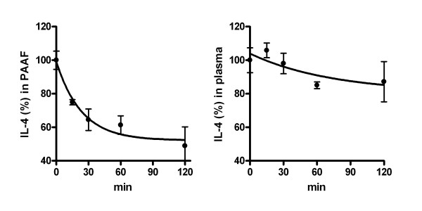
Background: Systemic inflammatory response syndrome is one of the major pathobiologic processes underlying severe acute pancreatitis and the degree of macrophage activation could be one of the factors that finally determine the severity of the disease. We evaluated the activation phenotype in peritoneal macrophages during the progression of an experimental model of acute pancreatitis induced in rats by intraductal administration of 5% sodium taurocholate and the effect of IL-4 and IL-13 to modulate this activation. Samples of pancreas, lung and adipose tissue as well as plasma were also obtained. In some animals IL4 and IL13 were injected 1 h after induction in order to modulate macrophage activation. The expressions of TNFalpha and Mannose Receptor, as indicators of classical and alternative macrophage activation, were evaluated. Levels of myeloperoxidase and plasma lipase were determined to evaluate the severity of the inflammatory process. The stability of IL-4 in ascitic fluid and plasma was evaluated.

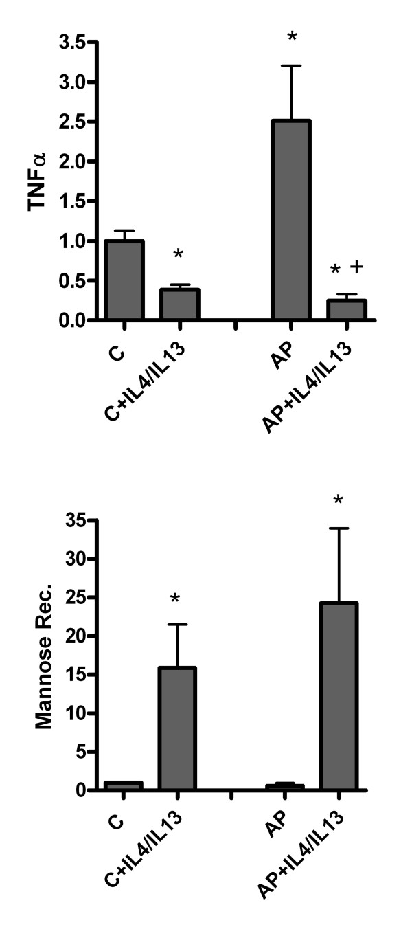
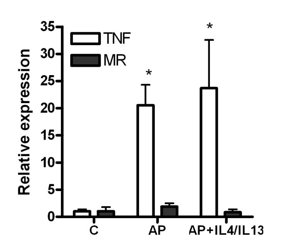
Results: Peritoneal macrophages showed a classical M1 activation clearly induced 3 h after pancreatitis induction and maintained until 18 h. Treatment with IL-4 and IL-13 reversed the activation of macrophages from a classical M1 to alternative M2 in vitro, but failed to modulate the response of peritoneal macrophages in vivo despite a reduction in inflammation was observed in lung and adipose tissue. Finally, IL-4 shows a short half-live in ascitic fluid when compared with plasma.

Conclusion: Peritoneal macrophages adopt a pro-inflammatory activation early during acute pancreatitis. Treatment with M2 cytokines could revert in vitro the pancreatitis-induced activation of macrophages but fails to modulate its activation in vivo. This treatment has only a moderate effect in reducing the systemic inflammation associated to acute pancreatitis. Hydrolytic enzymes presents in ascitic fluid could be involved in the degradation of cytokines, strongly reducing its utility to modulate peritoneal macrophages in pancreatitis.


Figures

Figure 1
Figure 1
Lipase and MPO activity in pancreas, lung and white adipose tissue (WAT) 3 and 8 hours after induction of pancreatitis. * = p < 0.05 vs. control.
Figure 2
Figure 2
Changes in the mRNA expression of TNFα and mannose receptor in peritoneal macrophages evaluated by RT-PCR. Results are expressed as increases with respect to the control group. * = p < 0.05 vs. control.
Figure 3
Figure 3
Reversibility of macrophage phenotype was observed on incubating peritoneal macrophages obtained 18 hours after pancreatitis induction and incubated with a mixture of IL-4 plus IL-13 for 24 hours. This treatment resulted in the inhibition of TNFα expression as well as the induction of Mannose receptor in both control and AP macrophages. Results are expressed as increases with respect to the control group. * = p < 0.05 vs. control; + = p < 0.05 vs AP.
Figure 4
Figure 4
In vivo administration of IL-4 plus IL-13 does not accomplish to reduce the macrophages activation in the early stages of pancreatitis. No changes were observed in TNFα or Mannose receptor expression in peritoneal macrophages. Results are expressed as increases with respect to the control group. * = p < 0.05 vs. control.
Figure 5
Figure 5
Effect of IL-4 and IL-13 administration on the progression of the early stages of inflammation. No changes were observed in lipase activity in plasma or in MPO levels in pancreas. By contrast, a significant inhibition of MPO in lung and white adipose tissue (WAT) indicates a reduction of the systemic inflammation. * = p < 0.05 vs. control; + = p < 0.05 vs AP.
Figure 6
Figure 6
Degradation of IL-4 in pancreatitis-associated ascitic fluid (left) and in plasma (right). PAAF or plasma from pancreatitis were pooled and overloaded with 25 pg/ml IL-4 and the concentration was measured at different time-points. In ascitic fluid, degradation of IL-4 was observed, with a half-life of 16.4 min. By contrast, half life in plasma was 51.1 min. Values are the mean of three different experiments.

Similar articles

Cited by

References

    1. Klöppel G, Maillet B. Pathology of acute and chronic pancreatitis. Pancreas. 1993;8:659–670. - PubMed
    1. Buter A, Imrie CW, Carter CR, Evans S, McKay CJ. Dynamic nature of early organ dysfunction determines outcome in acute pancreatitis. Br J Surg. 2003;89:298–302. doi: 10.1046/j.0007-1323.2001.02025.x. - DOI - PubMed
    1. Granger J, Remick D. Acute pancreatitis: models, markers, and mediators. Shock. 2005;24:45–51. doi: 10.1097/01.shk.0000191413.94461.b0. - DOI - PubMed
    1. Mikami Y, Takeda K, Shibuya K, et al. Do peritoneal macrophages play an essential role in the progression of acute pancreatitis in rats? Pancreas. 2003;27:253–260. doi: 10.1097/00006676-200310000-00011. - DOI - PubMed
    1. Closa D, Sabater L, Fernández-Cruz L, Prats N, Gelpí E, Roselló-Catafau J. Activation of alveolar macrophages in lung injury associated with experimental acute pancreatitis is mediated by the liver. Ann Surg. 1999;229:230–236. doi: 10.1097/00000658-199902000-00011. - DOI - PMC - PubMed

Publication types