Review
. 2009 Jul 31:7:79.
doi: 10.1186/1477-7827-7-79.

Vasodilator factors in the systemic and local adaptations to pregnancy

Affiliations
Review

Vasodilator factors in the systemic and local adaptations to pregnancy

Gloria Valdes et al. Reprod Biol Endocrinol. .

Abstract

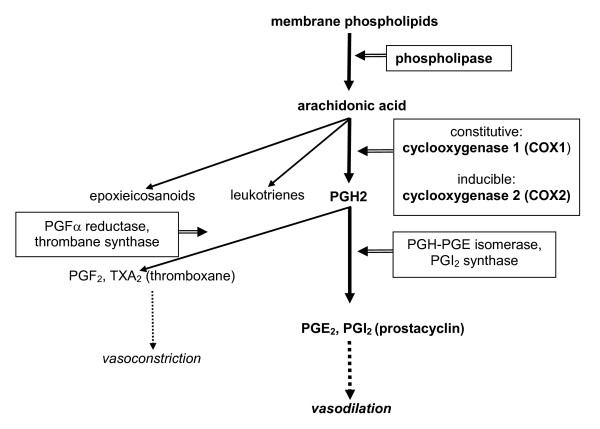
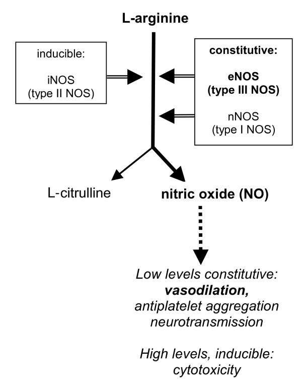
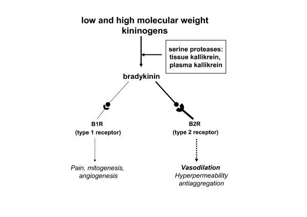
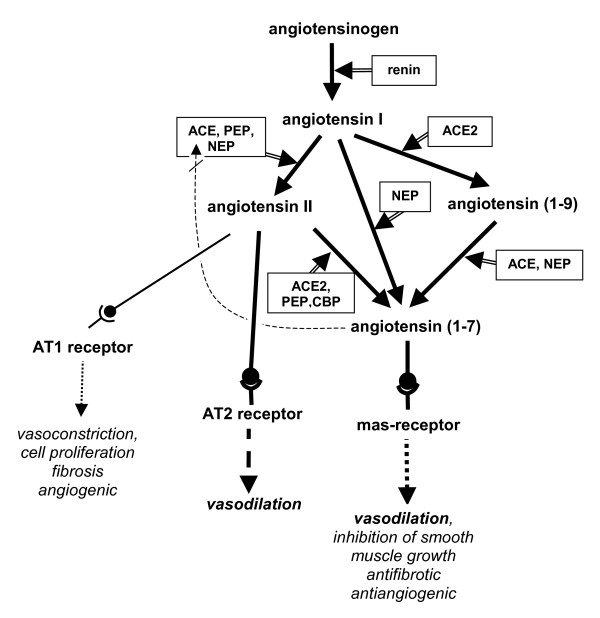
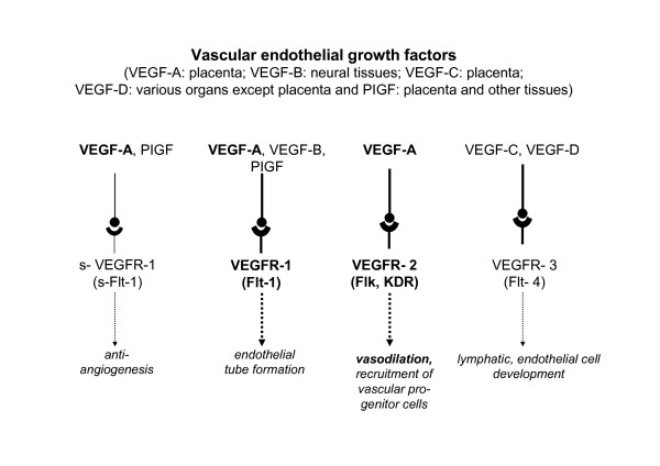
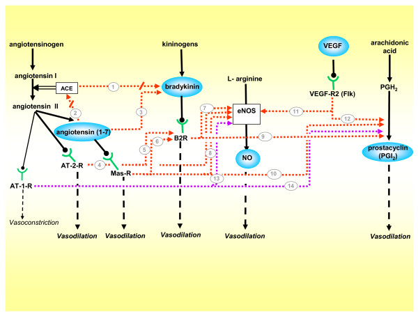
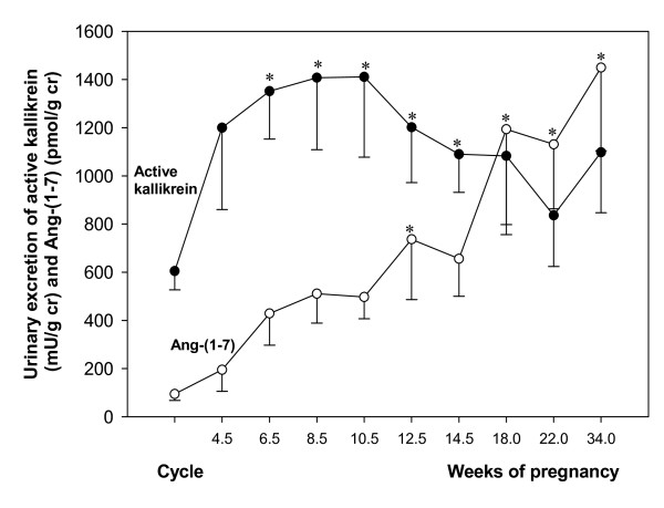
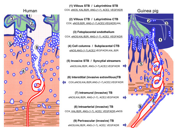
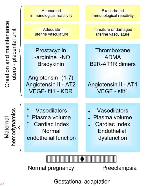
We postulate that an orchestrated network composed of various vasodilatory systems participates in the systemic and local hemodynamic adaptations in pregnancy. The temporal patterns of increase in the circulating and urinary levels of five vasodilator factors/systems, prostacyclin, nitric oxide, kallikrein, angiotensin-(1-7) and VEGF, in normal pregnant women and animals, as well as the changes observed in preeclamptic pregnancies support their functional role in maintaining normotension by opposing the vasoconstrictor systems. In addition, the expression of these vasodilators in the different trophoblastic subtypes in various species supports their role in the transformation of the uterine arteries. Moreover, their expression in the fetal endothelium and in the syncytiotrophoblast in humans, rats and guinea-pigs, favour their participation in maintaining the uteroplacental circulation. The findings that sustain the functional associations of the various vasodilators, and their participation by endocrine, paracrine and autocrine regulation of the systemic and local vasoactive changes of pregnancy are abundant and compelling. However, further elucidation of the role of the various players is hampered by methodological problems. Among these difficulties is the complexity of the interactions between the different factors, the likelihood that experimental alterations induced in one system may be compensated by the other players of the network, and the possibility that data obtained by manipulating single factors in vitro or in animal studies may be difficult to translate to the human. In addition, the impossibility of sampling the uteroplacental interface along normal pregnancy precludes obtaining longitudinal profiles of the various players. Nevertheless, the possibility of improving maternal blood pressure regulation, trophoblast invasion and uteroplacental flow by enhancing vasodilation (e.g. L-arginine, NO donors, VEGF transfection) deserves unravelling the intricate association of vasoactive factors and the systemic and local adaptations to pregnancy.


Figures

Figure 1
Figure 1
Synthesis of prostanoids and their respective vasodilator and vasoconstrictor actions.
Figure 2
Figure 2
L-arginine-nitric oxide (NO) pathway indicating the effects of NO according to its generating enzymes and tissue levels.
Figure 3
Figure 3
Kallikrein-kinin system, and the effects of bradykinin according to the type of receptor.
Figure 4
Figure 4
An updated version of the renin-angiotensin system and the different functions of the active peptides through their receptors.
Figure 5
Figure 5
Different forms of VEGF, their receptors and their respective functions.
Figure 6
Figure 6
Vasodilatory network, integrated by the five vasodilators included in this review, and their cognate interactions. While the majority of pathways are stimulatory, (1) ACE degrades bradykinin into inactive peptides, and diminishes its vasodilating effect[13], thus when (2) Ang-(1–7) competes with Ang I for ACE it indirectly potentiates bradykinin[16]. (3) Ang-(1–7) also enhances bradykinin activity[17]. (4) The AT-2-R stimulated by Angiotensin II activates eNOS, and (5) the B2R [23-25]. (6) The Mas-R activated by angiotensin-(1–7) stimulates the B2R[20], and (7,8) jointly with bradykinin activates eNOS[19](9,10). Activation of the B2R[13] and the Mas-R stimulates prostacyclin synthesis[21]. (11,12) The VEGF-R2 stimulates eNOS[23] and prostacyclin production[24]. Finally, the classical vasoconstrictor AT-1-R exerts in gestation a paradoxical vasodilatory effect by (13,14) stimulating the synthesis of NO and prostacyclin[95,96]. Stimulatory factors = blue balloons; stimulatory pathways = red arrows; inhibitory pathways = red arrows interrupted by oblique line; pathways exclusive to gestation = purple arrows.
Figure 7
Figure 7
Urinary excretion of angiotensin-(1–7) and active kallikrein in women in menstrual cycle and in 10 women with normotensive pregnancies[50,56].
Figure 8
Figure 8
Schematic representation of the placenta and fetoplacental junctional zone of humans and guinea-pigs. In order to highlight comparable structures, all fetal tissues are colored blue and green, all maternal structures red and brown. The main cellular and syncytial structures of both placentas are listed together with the reported expression of the vasodilator factors and enzymes. Factors are underlined when present in both species; isolated factors expressed only in one of the two species are lateralized to the presenting species. When the whole panel of factors is described in one of the species, this is depicted by a blue arrow pointing to the respective structures. Note that in the human an equivalent for periarterial trophoblast is not known. TB = trophoblasts; CTB = cytotrophoblasts; STB = syncytiotrophoblast.
Figure 9
Figure 9
Balance between immunological, morphological and vasoactive factors that determine an adequate or a defective adaptation to pregnancy, both at the level of the uteroplacental unit and the mother. For the purpose of this review emphasis has been given to the local and systemic hemodynamic adaptations.

Similar articles

Cited by

References

    1. Kaufmann P, Black S, Huppertz B. Endovascular trophoblast invasion: implications for the pathogenesis of intrauterine growth retardation and preeclampsia. Biol Reprod. 2003;69:1–7. - PubMed
    1. Red-Horse K, Zhou Y, Genbacev O, Prakobphol A, Foulk R, McMaster M, Fisher SJ. Trophoblast differentiation during embryo implantation and formation of the maternal-fetal interface. J Clin Invest. 2004;114:744–754. - PMC - PubMed
    1. Kaufmann P, Mayhew TM, Charnock-Jones DS. Aspects of human fetoplacental vasculogenesis and angiogenesis. II. Changes during normal pregnancy. Placenta. 2004;25:114–126. - PubMed
    1. Reynolds LP, Redmer DA. Angiogenesis in the placenta. Biol Reprod. 2001;64:1033–1040. - PubMed
    1. Hytten F. Blood volume changes in normal pregnancy. Clin Haematol. 1985;14:601–612. - PubMed

Publication types

LinkOut - more resources