

Drosophila cortex and neuropile glia influence secondary axon tract growth, pathfinding, and fasciculation in the developing larval brain
- PMID: 19646433
- PMCID: PMC2776086
- DOI: 10.1016/j.ydbio.2009.07.035
Drosophila cortex and neuropile glia influence secondary axon tract growth, pathfinding, and fasciculation in the developing larval brain
Abstract
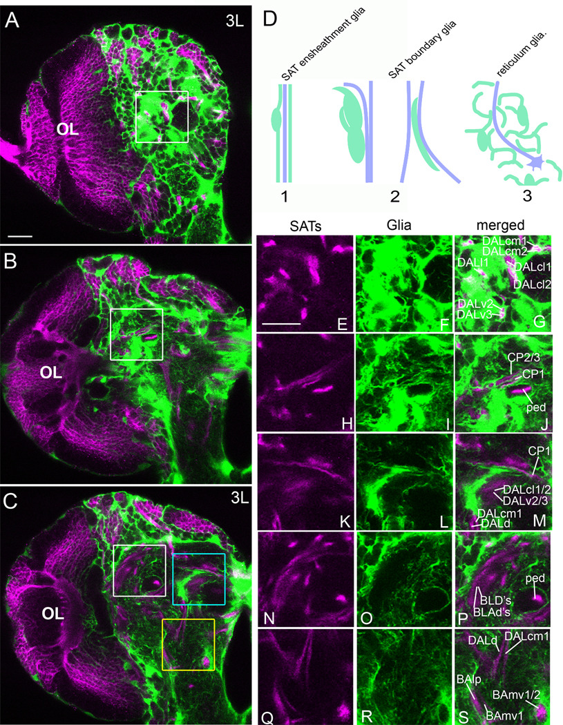
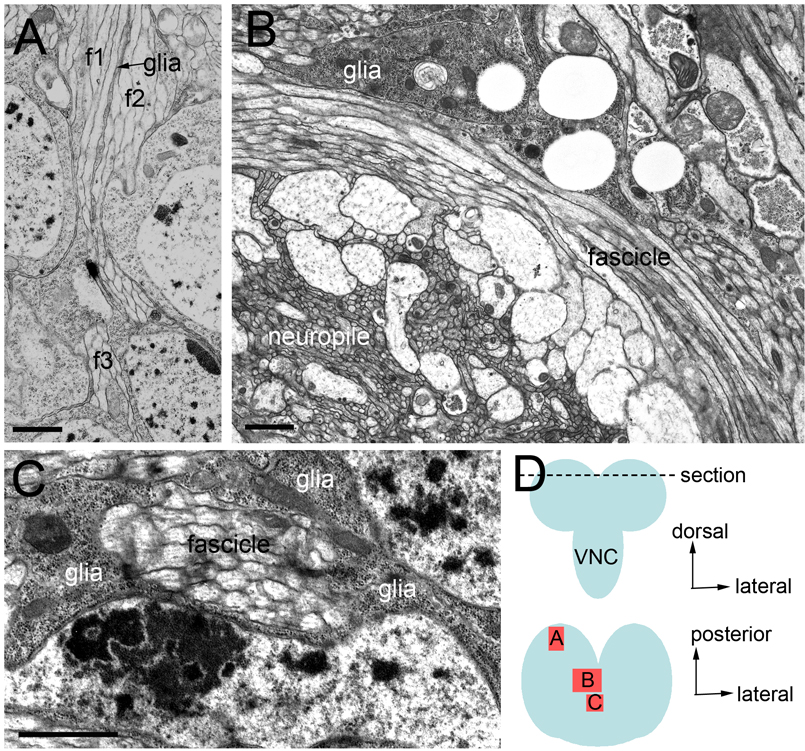
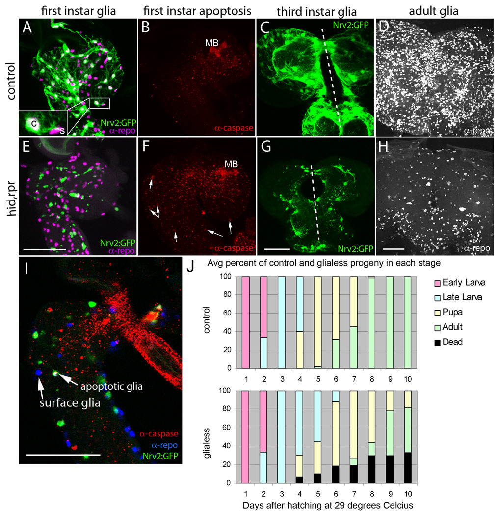
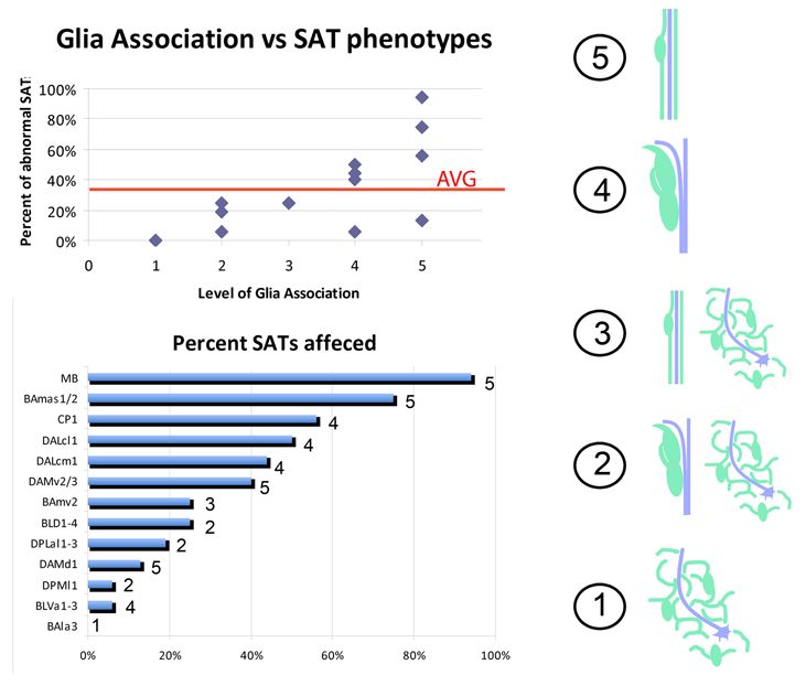
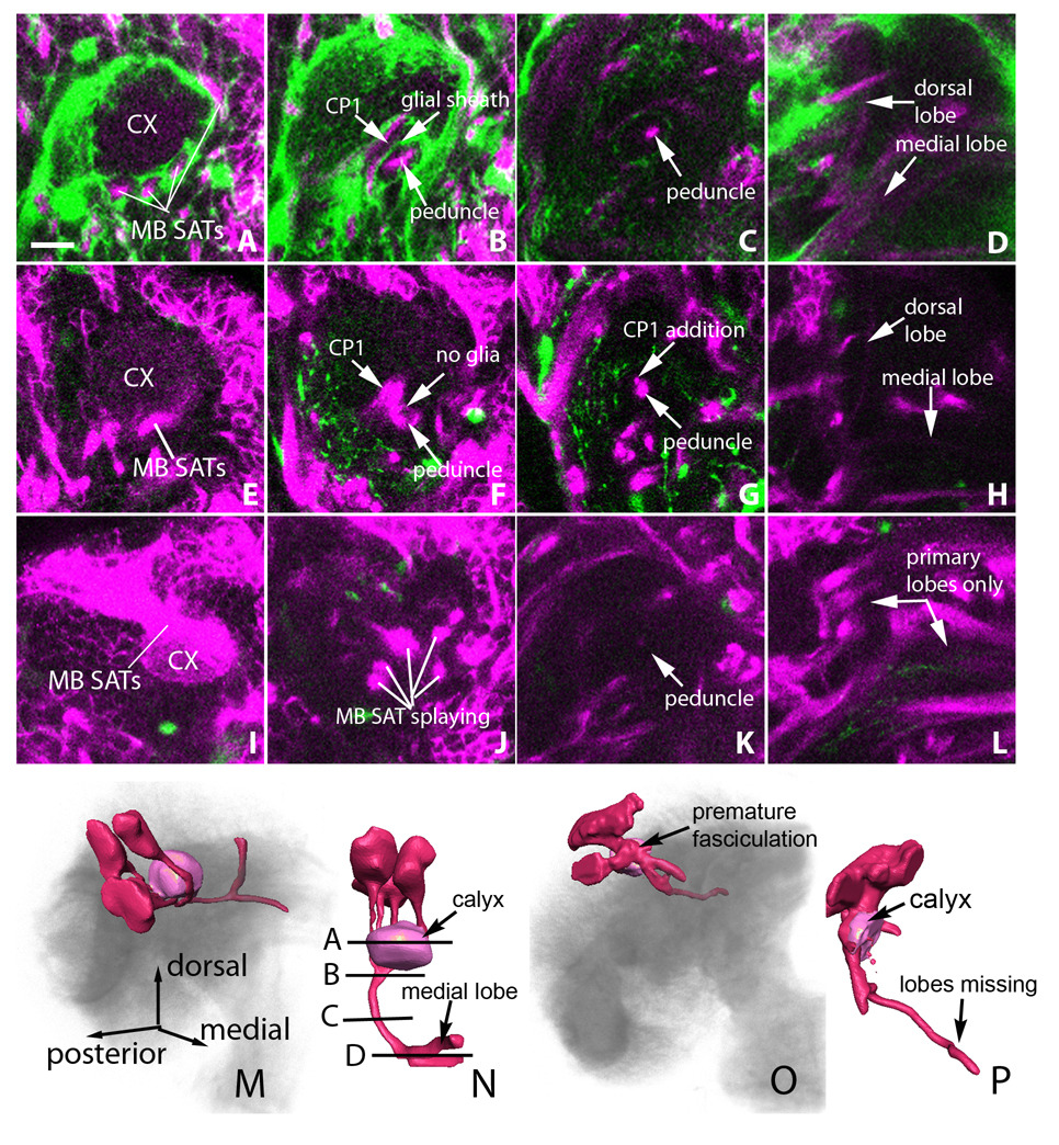
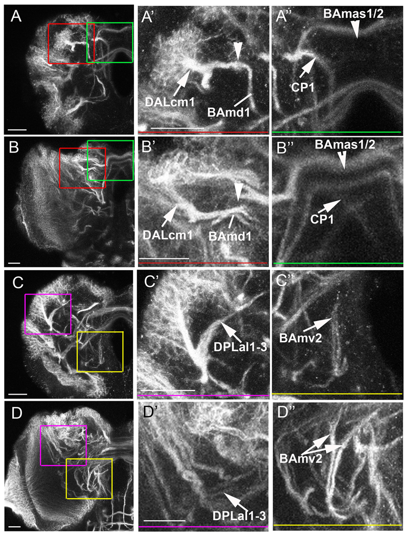
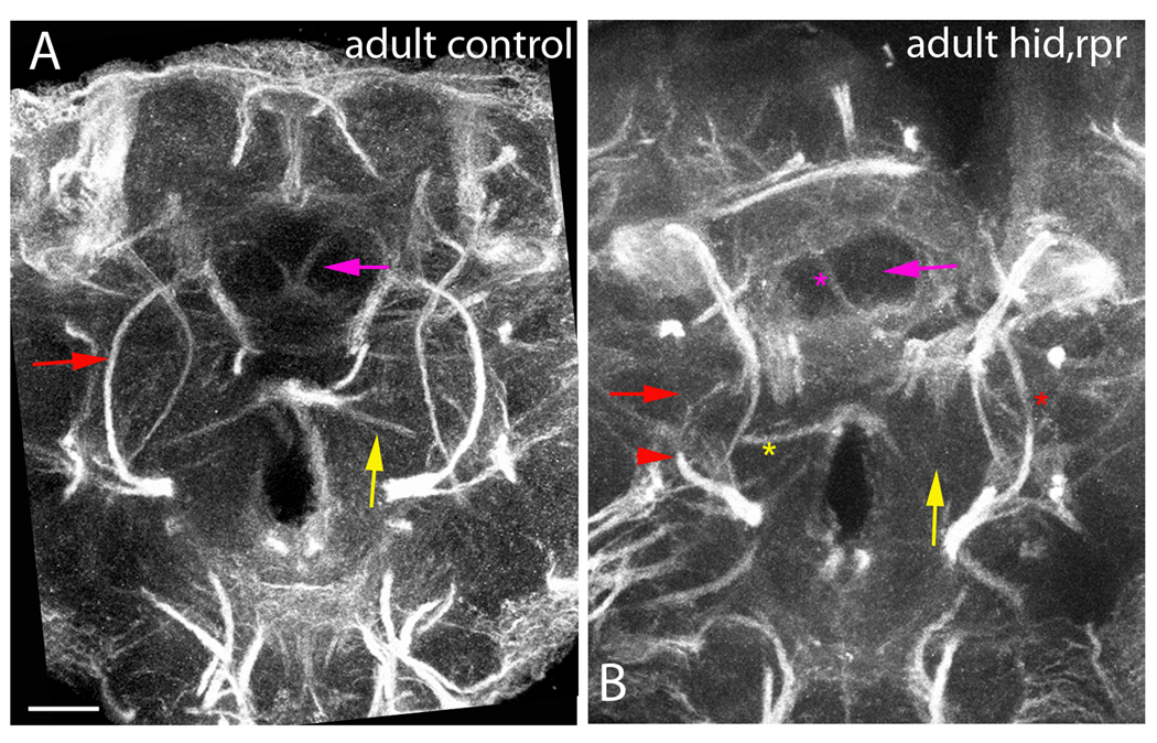
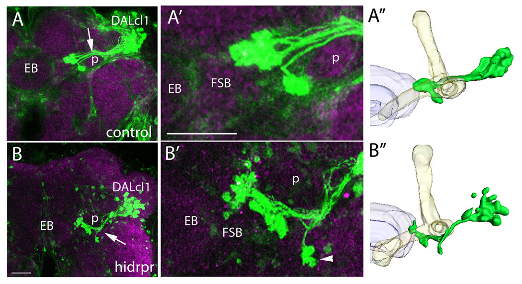
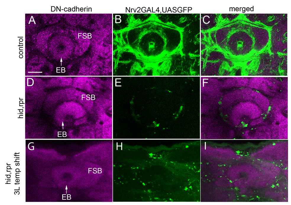
Glial cells play important roles in the developing brain during axon fasciculation, growth cone guidance, and neuron survival. In the Drosophila brain, three main classes of glia have been identified including surface, cortex, and neuropile glia. While surface glia ensheaths the brain and is involved in the formation of the blood-brain-barrier and the control of neuroblast proliferation, the range of functions for cortex and neuropile glia is less well understood. In this study, we use the nirvana2-GAL4 driver to visualize the association of cortex and neuropile glia with axon tracts formed by different brain lineages and selectively eliminate these glial populations via induced apoptosis. The larval central brain consists of approximately 100 lineages. Each lineage forms a cohesive axon bundle, the secondary axon tract (SAT). While entering and traversing the brain neuropile, SATs interact in a characteristic way with glial cells. Some SATs are completely invested with glial processes; others show no particular association with glia, and most fall somewhere in between these extremes. Our results demonstrate that the elimination of glia results in abnormalities in SAT fasciculation and trajectory. The most prevalent phenotype is truncation or misguidance of axon tracts, or abnormal fasciculation of tracts that normally form separate pathways. Importantly, the degree of glial association with a given lineage is positively correlated with the severity of the phenotype resulting from glial ablation. Previous studies have focused on the embryonic nerve cord or adult-specific compartments to establish the role of glia. Our study provides, for the first time, an analysis of glial function in the brain during axon formation and growth in larval development.
Figures
References
-
- Ashburner M. A Laboratory Handbook. Cold Spring Harbor Laboratory Press; 1989.
-
- Bach EA, Ekas LA, Ayala-Camargo A, Flaherty MS, Lee H, Perrimon N, Baeg GH. GFP reporters detect the activation of the Drosophila JAK/STAT pathway in vivo. Gene Expr Patterns. 2007;7:323–331. - PubMed
-
- Bainton RJ, Tsai LT, Schwabe T, DeSalvo M, Gaul U, Heberlein U. Moody encodes two GPCRs that regulate cocaine behaviors and blood-brain barrier permeability in Drosophila. Cell. 2005;123:145–156. - PubMed
-
- Baumann PM, Oland LA, Tolbert LP. Glial cells stabilize axonal protoglomeruli in the developing olfactory lobe of the moth Manduca sexta. J Comp Neurol. 1996;373:118–128. - PubMed
Publication types
MeSH terms
Substances
Grants and funding
LinkOut - more resources
Full Text Sources
Molecular Biology Databases
Research Materials
