. 2009 Sep 15;19(17):1421-8.
doi: 10.1016/j.cub.2009.07.018. Epub 2009 Jul 30.

Mechanosensing through cooperative interactions between myosin II and the actin crosslinker cortexillin I

Affiliations

Mechanosensing through cooperative interactions between myosin II and the actin crosslinker cortexillin I

Yixin Ren et al. Curr Biol. .

Abstract

Background: Mechanosensing governs many processes from molecular to organismal levels, including during cytokinesis where it ensures successful and symmetrical cell division. Although many proteins are now known to be force sensitive, myosin motors with their ATPase activity and force-sensitive mechanical steps are well poised to facilitate cellular mechanosensing. For a myosin motor to experience tension, the actin filament must also be anchored.

Results: Here, we find a cooperative relationship between myosin II and the actin crosslinker cortexillin I where both proteins are essential for cellular mechanosensory responses. Although many functions of cortexillin I and myosin II are dispensable for cytokinesis, all are required for full mechanosensing. Our analysis demonstrates that this mechanosensor has three critical elements: the myosin motor where the lever arm acts as a force amplifier, a force-sensitive bipolar thick-filament assembly, and a long-lived actin crosslinker, which anchors the actin filament so that the motor may experience tension. We also demonstrate that a Rac small GTPase inhibits this mechanosensory module during interphase, allowing the module to be primarily active during cytokinesis.

Conclusions: Overall, myosin II and cortexillin I define a cellular-scale mechanosensor that controls cell shape during cytokinesis. This system is exquisitely tuned through the enzymatic properties of the myosin motor, its lever arm length, and bipolar thick-filament assembly dynamics. The system also requires cortexillin I to stably anchor the actin filament so that the myosin motor can experience tension. Through this cross-talk, myosin II and cortexillin I define a cellular-scale mechanosensor that monitors and corrects shape defects, ensuring symmetrical cell division.


Figures

Fig. 1
Fig. 1
Myosin-II lever arm length determines the pressure-threshold-dependent behavior of the cellular mechanosensory response. (A) Representative micrographs showing a positive response to applied pressure. The cell is a myoII: Cit-ΔBLCBS;GFP-tubulin. Top panels, DIC images. Lower panels, fluorescence images. Left panels, cell before aspiration; right panels, cell during aspiration. The centrosomes are visible and Cit-ΔBLCBS accumulates at the micropipette (arrow). This cell is one of the positive responses of ΔBLCBS at 0.39 nN/µm2 pressure. Scale bar, 10 µm. (B) Micrograph of a mitotic cell expressing wild type GFP-myosin-II, showing a response. The intensity of the cortex inside the micropipette (Ip) and the opposite cortex (Io) were measured. The Ip/Io ratio was calculated and the log transform used for analysis. (C) Cartoon comparing wild type, 2xELC, ΔBLCBS, and S456L motors. Blue/pink, motor domain; yellow, essential light chain; red, regulatory light chain. (D) Graph shows the dependency of the fraction of responses on the applied pressure. Frequency histograms of each dataset and a second graph showing the overall average magnitudes (±SEMs) are provided in Fig. S3 (see Experimental Procedures also). At 0.15 nN/µm2 pressure, 2xELC is more responsive than wild type, S456L, or ΔBLCBS myosins (Student's t –test: P<0.01). Wild type and 2xELC myosin-II are more responsive than ΔBLCBS at 0.39 and 0.64 nN/µm2 pressure (Student’s t –test: P<0.01).
Fig. 2
Fig. 2
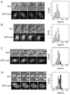
The mechanosensitive localization of cortexillin-I requires myosin-II. Example time series (times in s) of DIC and fluorescent images are shown for (A) a myoII:GFP-cortI cell aspirated with 0.30 nN/µm2 of pressure; (B) a myoII:GFP-cortI;dynhp cell aspirated with 0.21–0.28 nN/µm2 of pressure; (C) a myoII:GFP-cortI;S456L cell aspirated with 0.26 nN/µm2 of pressure; and (D) a myoII:GFP-cortI; myosin (rescue) cell aspirated with 0.45 nN/µm2 of pressure. Frequency histograms show measurements from all cells measured for each genotype. As described in the Experimental Procedures, the dark grey bars of the histograms indicate positive responses, while light grey bars indicate negative responses. Statistical analysis indicated that the myoII:GFP-cortI and myoII:GFP-cortI;myoII strains are statistically distinct (Student’s t –test: P<0.001). Scale bar, 10 µm.
Fig. 3
Fig. 3
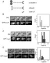
The mechanosensitive localization of myosin-II requires cortexillin-I. (A) Wild type cortexillin-I, ΔNcortl and cortI CT were tested for their ability to restore mechanosensory responses. All three proteins rescue cytokinesis [24, 25]. Example time series (times in s) of DIC and fluorescence images are shown for (B) a cortI:GFP-myoII cell; (C) a cortI: GFP-myoII;RFP-cortI (full-length cortexillin-I) cell; and (D) a cortI: GFP-myoII;RFP-ΔNcortl cell. Frequency histograms show measurements from all cells measured for each genotype. Statistical analysis indicated that the cortI: GFP-myoII;RFP-tub and cortI: GFP-myoII;RFP-cortI strains are statistically distinct (Student’s t –test: P<0.0001). Scale bar, 10 µm.
Fig. 4
Fig. 4
Single molecule analysis of cortexillin-I-actin interactions. (A) Cartoon depicts the geometry of the experimental set up. GFP-cortexillin-I is anchored to the substrate through the GFP using anti-GFP antibodies. An actin dumbbell is steered into position using a dual beam optical trap. (B) An example trace showing the bead position (top) and the cross-correlation of the fluctuations of the two beads (bottom) holding the actin dumbbell. (C) Dwell time distribution showing the distribution of bound life-times. The mean τ is 550 ms (± 40 ms, n = 776 events). Errors are standard errors from fitting bootstrap sampled datasets.
Fig. 5
Fig. 5
RacE is the cell cycle stage specificity factor that determines when myosin-II and cortexillin-I can redistribute in response to mechanical strain. Example time series (times in s) of DIC and fluorescence images are shown for (A) a mitotic RacE: GFP-myoII cell; (B) an interphase RacE: GFP-myoII cell; (C) an interphase RacE: GFP-cortI cell; and (D) an interphase RacE: mCh-RacE;GFP-myoII cell. Frequency histograms show measurements from all cells measured for each genotype. Scale bar, 10 µm.
Fig. 6
Fig. 6
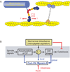
Mechanosensory cell shape control system. (A) Cartoon depicts the mechanical circuit between myosin-II and cortexillin-I that mediates mechanosensing. Because tension is required to balance the myosin power stroke, which generates a force (F) on the actin filament, cortexillin-I likely anchors the actin filament, providing the tension (T) needed to increase the strongly bound state time (τs). This cross-communication between myosin-II and cortexillin-I stabilizes each protein on the actin, promoting their accumulation. This stabilization also appears to provide feedback on myosin-II thick filament assembly, allowing thick filaments to form, a requisite for accumulation. (B) This mechanosensory system ensures successful high fidelity cytokinesis. Mechanical perturbation halts cytokinesis during early stages of cytokinesis and triggers accumulation of myosin-II and cortexillin-I to the site of mechanical deformation during all stages of cytokinesis. Cooperative interactions between myosin-II and cortexillin-I define the cellular-scale mechanosensor.

Similar articles

Cited by

References

    1. Orr AW, Helmke BP, Blackman BR, Schwartz MA. Mechanisms of mechanotransduction. Dev. Cell. 2006;10:11–20. - PubMed
    1. Effler JC, Kee Y-S, Berk JM, Tran MN, Iglesias PA, Robinson DN. Mitosis-specific mechanosensing and contractile protein redistribution control cell shape. Curr. Biol. 2006;16:1962–1967. - PMC - PubMed
    1. Engler AJ, Sen S, Sweeney HL, Discher DE. Matrix elasticity directs stem cell lineage specification. Cell. 2006;126:677–689. - PubMed
    1. Martinac B. Mechanosensitive ion channels: molecules of mechanotransduction. J. Cell Sci. 2004;117:2449–2460. - PubMed
    1. Balaban NQ, Schwartz US, Riveline D, Goichberg P, Tzur G, Sabanay I, Mahalu D, Safran S, Bershadsky AD, Addadi L, Geiger B. Force and focal adhesion assembly: a close relationship studied using elastic micropatterned substrates. Nat. Cell Biol. 2001;3:466–472. - PubMed

Publication types

MeSH terms

Substances