. 2009 Aug 12;17(8):1075-81.
doi: 10.1016/j.str.2009.06.012. Epub 2009 Jul 30.

Characterizing the initial encounter complex in cadherin adhesion

Affiliations

Characterizing the initial encounter complex in cadherin adhesion

Sanjeevi Sivasankar et al. Structure. .

Abstract

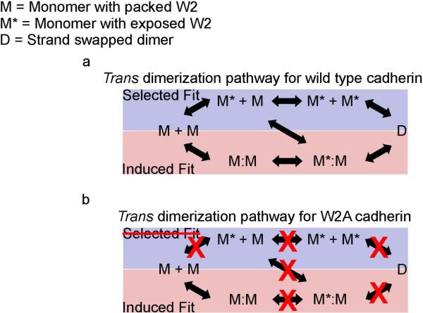
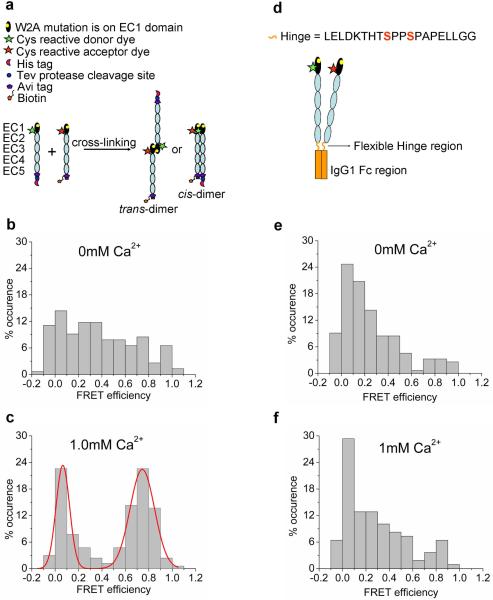
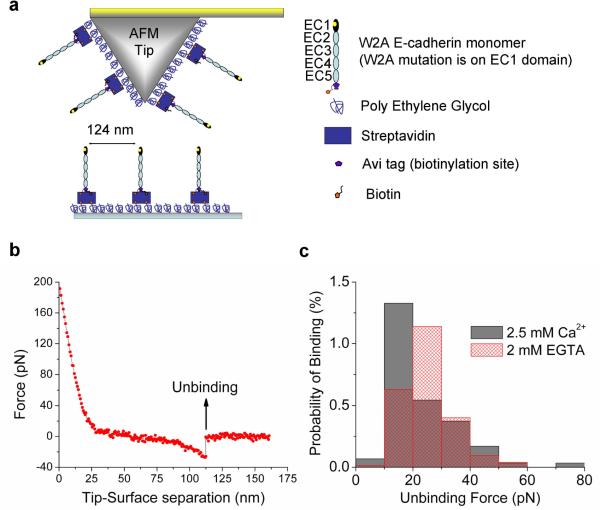
Cadherins are Ca(2+)-dependent cell-cell adhesion proteins with an extracellular region of five domains (EC1 to EC5). Adhesion is mediated by "strand swapping" of a conserved tryptophan residue in position 2 between EC1 domains of opposing cadherins, but the formation of this structure is not well understood. Using single-molecule fluorescence resonance energy transfer and single-molecule force measurements with the atomic force microscope, we demonstrate that cadherins initially interact via EC1 domains without swapping tryptophan-2 to form a weak Ca(2+) dependent initial encounter complex that has 25% of the bond strength of a strand-swapped dimer. We suggest that cadherin dimerization proceeds via an induced fit mechanism where the monomers first form a tryptophan-2 independent initial encounter complex and then undergo subsequent conformational changes to form the final strand-swapped dimer.


Figures

Figure 1
Figure 1. Alternative pathways for the formation of strand-exchange dimers in wild type cadherin and W2A cadherin
Figure 1a has been adopted from (Miloushev et al., 2008) (a)Wild type cadherin: In the minimal selected-fit pathway (blue box), a population of cadherin monomers (M) adopt an active conformation (M*) by exposing their W2 residues. Collisions between activated cadherin monomers result in the formation of a stand swapped dimer (D). In the minimal induced-fit pathway (pink box), two cadherin monomers with intramolecularly packed W2 first form an initial encounter complex (M:M). A conformational change then results in the swapping of W2 residues and the formation of a strand-swapped dimer. (b)W2A cadherin: Mutating the W2 residue eliminates the selected-fit pathway for formation of a strand-swapped dimer. Interactions between individual W2 mutants would occur only if the cadherins dimerize via an induced-fit mechanism. Since the interacting W2 mutant cadherins cannot proceed to form a strand swapped dimer, they stall at the initial encounter complex which can be detected and characterized using single molecule techniques.
Figure 2
Figure 2. Characterizing the structure of the initial encounter complex using single molecule FRET
(a) Steps involved in cross-linking EC1 labeled W2A cadherin monomers. W2A cadherin monomers engineered with a His-affinity tag or a biotin-tag were labeled with a donor Cy3 fluorophore or acceptor Alexa-647 fluorophore respectively at N20C. Donor- and acceptor-labeled cadherins were cross-linked in solution using amine reactive BS3 cross-linker. Cross-linked dimer yields in 0mM Ca2+ and 1mM Ca2+ were 45% and 20%, respectively. The higher cross-linking yield in 0mM Ca2+ was attributed to the high levels of nonspecific interaction of W2A cadherin in the absence of Ca2+ (see below). (b) Histogram of FRET efficiencies for W2A cadherin dimers cross-linked at 0mM Ca2+ shows a broad distribution of FRET efficiencies without a pronounced peak. This distribution arises because of the nonspecific interaction of the EC1 domains of the W2A mutants in the absence of Ca2+. (c) Histogram of FRET efficiencies for W2A cadherin dimers cross-linked at 1mM Ca2+ shows a pronounced peak at a FRET efficiency of 0.74 with 84% of the co-localized fluorescence spots have FRET values greater than 0.5. A FRET value of 0.74 corresponds to a distance of approximately 4nm between the donor and acceptor fluorophores and arises due to the interaction of the EC1 domains in the initial encounter complex. (d) Schematic of W2A cadherin-Fc dimer construct engineered by fusing the COOH terminus of the cadherin extracellular region to the Fc domain of human IgG1. Engineered cysteines (N20C) on the EC1 domains of the cadherins were dual labeled with donor and acceptor fluorophores. The cysteine residues in the core hinge region had been mutated to serines to prevent nonspecific labeling of the Fc dimer. (e-f) Histogram of FRET efficiencies for W2A cadherin-Fc dimers cross-linked at 0mM and 1mM Ca2+. W2A cadherin-Fc shows a broad distribution of FRET efficiencies without a pronounced peak due to the nonspecific interaction of the EC1 domains when the W2A cadherin are constrained in a cis orientation.
Figure 3
Figure 3. Characterizing the bond-strength of the initial encounter complex using single molecule AFM force measurements
(a) Schematic of Atomic Force Microscope (AFM) tip and substrate functionalized with biotinylated W2A cadherin monomers for single molecule force measurements. The tip and surface were functionalized with Poly Ethylene Glycol (PEG) linkers, some of which were decorated with Streptavidin. Biotinylated W2A cadherin monomers were bound to Streptavidin on the tip and surface; flexible PEG linkers enable unhindered interactions between opposing cadherins during tip-substrate encounters. (b) A typical force curve showing the unbinding of a single W2A cadherin molecule. The tip and the substrate decorated with cadherins were bought into contact so that cadherins on the tip and substrate formed an adhesive complex. When the tip was withdrawn from the substrate a force was exerted and above a critical force the adhesive complex ruptured. Forces were measured 2937 times in 2.5mM Ca2+ yielding 75 binding events and 7467 times in 2mM EGTA yielding 173 binding events. (c) Histogram of W2A cadherin monomer binding events measured in 2.5mM Ca2+ (solid gray bars) and 2mM EGTA (hatched red bars). The binding events measured in 2.5mM Ca2+ were fitted to a Gaussian distribution with a peak force of 17 pN ± 10pN. The probability of binding did not change in the absence of Ca2+ (hatched red bar).

Comment in

References

    1. Aitken CE, Marshall RA, Pulglisi JD. An oxygen scavenging system for improvement of dye stability in single-molecule fluorescence experiments. Biophys. J. 2008;94:1826–1835. - PMC - PubMed
    1. Al-Amoudi A, Diez DC, Betts MJ, Frangakis AS. The molecular architecture of cadherins in native epidermal desmosomes. Nature. 2007;450:832–U838. - PubMed
    1. Boggon TJ, Murray J, Chappuis-Flament S, Wong E, Gumbiner BM, Shapiro L. C-cadherin ectodomain structure and implications for cell adhesion mechanisms. Science. 2002;296:1308–1313. - PubMed
    1. Chien YH, Jiang N, Li F, Zhang F, Zhu C, Leckband D. Two stage cadherin kinetics require multiple extracellular domains but not the cytoplasmic region. J. Biol. Chem. 2008;283:1848–1856. - PMC - PubMed
    1. Chitaev NA, Troyanovsky SM. Adhesive but not lateral E-cadherin complexes require calcium and catenins for their formation. J. Cell Biol. 1998;142:837–846. - PMC - PubMed

Publication types

MeSH terms

LinkOut - more resources