. 2010 Jan;48(1):181-90.
doi: 10.1016/j.yjmcc.2009.07.020. Epub 2009 Jul 30.

Augmented potassium current is a shared phenotype for two genetic defects associated with familial atrial fibrillation

Affiliations

Augmented potassium current is a shared phenotype for two genetic defects associated with familial atrial fibrillation

Robert L Abraham et al. J Mol Cell Cardiol. 2010 Jan.

Abstract

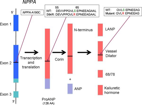
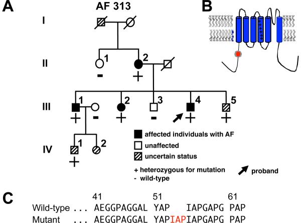
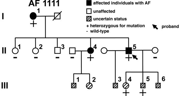
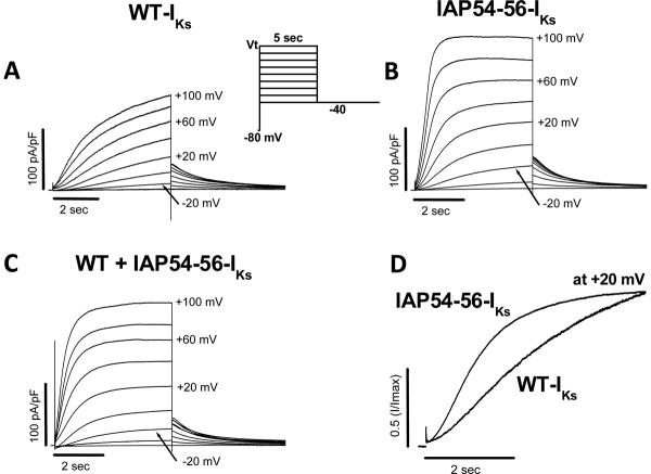
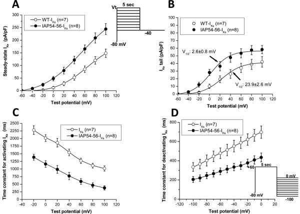
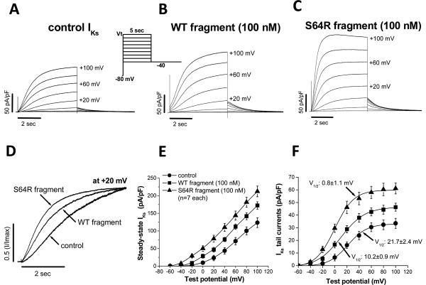
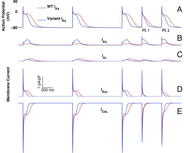
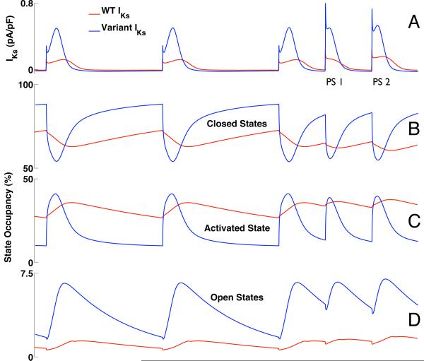
Mutations in multiple genes have been implicated in familial atrial fibrillation (AF), but the underlying mechanisms, and thus implications for therapy, remain ill-defined. Among 231 participants in the Vanderbilt AF Registry, we found a mutation in KCNQ1 (encoding the alpha-subunit of slow delayed rectifier potassium current [I(Ks)]) and separately a mutation in natriuretic peptide precursor A (NPPA) gene (encoding atrial natriuretic peptide, ANP), both segregating with early onset lone AF in different kindreds. The functional effects of these mutations yielded strikingly similar I(Ks) "gain-of-function." In Chinese Hamster Ovary (CHO) cells, coexpression of mutant KCNQ1 with its ancillary subunit KCNE1 generated approximately 3-fold larger currents that activated much faster than wild-type (WT)-I(Ks). Application of the WT NPPA peptide fragment produced similar changes in WT-I(Ks), and these were exaggerated with the mutant NPPA S64R peptide fragment. Anantin, a competitive ANP receptor antagonist, completely inhibited the changes in I(Ks) gating observed with NPPA S64R. Computational simulations identified accelerated transitions into open states as the mechanism for variant I(Ks) gating. Incorporating these I(Ks) changes into computed human atrial action potentials (AP) resulted in 37% shortening (120 vs. 192 ms at 300 ms cycle length), reflecting loss of the phase II dome which is dependent on L-type calcium channel current. We found striking functional similarities due to mutations in KCNQ1 and NPPA genes which led to I(Ks) "gain-of-function", atrial AP shortening, and consequently altered calcium current as a common mechanism between diverse familial AF syndromes.


Figures

Figure 1
Figure 1
NPPA post-transcriptional processing. The initial 151-peptide product, PreproANP, is further processed into ANP and a collection of other protein fragments, including the N-terminal proANP `vessel dilator' peptide fragment. The NPPA-A190C mutation leading to PreproANPS64R propagates into this vessel dilator peptide fragment. The amino acid sequences of the WT and mutant fragments are shown (inset). (Figure modified from Veseley, 2001 [19].)
Figure 2
Figure 2
KCNQ1 mutation identified in a kindred with familial atrial fibrillation (AF). (A) Family pedigree of Vanderbilt AF 313. Solid symbols denote AF and striped symbols individuals of uncertain phenotype. Male subjects are shown as squares and female subjects as circles. The proband (arrow) and the presence (+) or absence (-) of the KCNQ1-IAP54-56 indel are indicated. (B) Location of the variant in the KCNQ1 protein. (C) Portion of the KCNQ1 N-terminus amino acid wild-type and mutant sequence.
Figure 3
Figure 3
NPPA mutation identified in a kindred with familial AF. Shown is the family pedigree of Vanderbilt AF 1111. Symbols as in Figure 1A.
Figure 4
Figure 4
Comparison of currents recorded from (A) wild-type (WT)-IKs (KCNQ1 + KCNE1), (B) IAP54-56-IKs (KCNQ1-IAP54-56 + KCNE1) and (C) co-expression of the WT-IKs + IAP54-56-IKs channels in CHO cells during 5-sec depolarization steps from a holding potential of -80 mV to test potentials in the range -40 to +100 mV followed by repolarization to -40 mV (voltage protocol shown in inset). (D) Comparison of activation of WT and IAP54-56-IKs. Traces obtained at +20 mV are redrawn from (A) and (B) at +20 mV.
Figure 5
Figure 5
Current/voltage (I/V) relationships for WT-IKs and mutant (IAP54-56-IKs) channels. (A) Activating IKs as a function of voltage, normalized to cell surface area and measured at the end of a 5-sec depolarizing pulse. (B) Normalized tail current amplitude at -40 mV as a function of depolarizing potential. Fits to Boltzmann distributions are shown. (C) Time constant of IKs activation as a function of voltage. Time constants of IKs deactivation as a function of voltage are shown in (D).
Figure 6
Figure 6
Effects of WT NPPA peptide fragment and mutant NPPA peptide fragment-S64R on IKs. (A) control IKs. (B) Increased IKs on exposure to WT fragment. (C) Accelerated activation and further increase of IKs with S64R fragment. (D) Superimposition of activating IKs at +20 mV from panels (A), (B), and (C). (E) Steady state IKs normalized to cell surface area (measured at the end of a 5-sec depolarizing pulse). (F) Normalized tail current amplitude at -40 mV as a function of depolarizing potential. Fits to Boltzmann distributions are shown.
Figure 7
Figure 7
Simulated double premature atrial depolarizations (400 ms coupling intervals) after a twelve-pulse pacing train (1000 ms cycle length). Data from simulations that include WT-IKs are shown in red, and those incorporating variant IKs, based on IAP54-56, are in blue (A). The first atrial premature stimulus (PS1) simulated with variant IKs was shorter than that for WT-IKs (126 vs. 193 ms). AP durations for the second premature stimulus (PS2) were 138 ms and 243 ms for variant IKs and WT-IKs, respectively. In both simulations, PS1 exhibited loss of the dome associated with membrane repolarization. However, with WT-IKs the dome was restored with PS2 while it remained absent with variant IKs. Changes in individual membrane currents are shown in (B) IKs; (C) IKr; (D) IKur; and (E) ICa-L.
Figure 8
Figure 8
IKs and channel state occupancy during the simulation of Figure 7 (1000 ms pacing train followed by double premature atrial depolarizations with 400ms coupling interval). WT-IKs is shown in red; variant IKs is in blue (A). Variant IKs exhibits much more dynamic variation in state occupancy during activation as channels transition from the closed states (B) through the activated state (C) into the open states (D). `Closed States' refers to the sum of closed states R1 and R2; `Open States' (D) refers to the sum of open states O1 and O2 (see Figure S-1A of the online data supplement).

References

    1. Go AS, Hylek EM, Phillips KA, Chang Y, Henault LE, Selby JV, et al. Prevalence of diagnosed atrial fibrillation in adults: national implications for rhythm management and stroke prevention: the AnTicoagulation and Risk Factors in Atrial Fibrillation (ATRIA) Study. JAMA. 2001;285(18):2370–5. - PubMed
    1. Lloyd-Jones DM, Wang TJ, Leip EP, Larson MG, Levy D, Vasan RS, et al. Lifetime risk for development of atrial fibrillation: the Framingham Heart Study. Circulation. 2004;110(9):1042–6. - PubMed
    1. Wolf PA, Abbott RD, Kannel WB. Atrial fibrillation as an independent risk factor for stroke: the Framingham Study. Stroke. 1991;22(8):983–8. - PubMed
    1. Benjamin EJ, Wolf PA, D'Agostino RB, Silbershatz H, Kannel WB, Levy D. Impact of atrial fibrillation on the risk of death: the Framingham Heart Study. Circulation. 1998;98(10):946–52. - PubMed
    1. Gersh BJ, Tsang TS, Seward JB. The changing epidemiology and natural history of nonvalvular atrial fibrillation: clinical implications. Trans Am Clin Climatol Assoc. 2004;115:149–60. - PMC - PubMed

Publication types