Review
. 2010 Feb;1800(2):80-95.
doi: 10.1016/j.bbagen.2009.07.017. Epub 2009 Jul 30.

The hexosamine signaling pathway: O-GlcNAc cycling in feast or famine

Affiliations
Review

The hexosamine signaling pathway: O-GlcNAc cycling in feast or famine

John A Hanover et al. Biochim Biophys Acta. 2010 Feb.

Abstract

The enzymes of O-GlcNAc cycling couple the nutrient-dependent synthesis of UDP-GlcNAc to O-GlcNAc modification of Ser/Thr residues of key nuclear and cytoplasmic targets. This series of reactions culminating in O-GlcNAcylation of targets has been termed the hexosamine signaling pathway (HSP). The evolutionarily ancient enzymes of O-GlcNAc cycling have co-evolved with other signaling effecter molecules; they are recruited to their targets by many of the same mechanisms used to organize canonic kinase-dependent signaling pathways. This co-recruitment of the enzymes of O-GlcNAc cycling drives a binary switch impacting pathways of anabolism and growth (nutrient uptake) and catabolic pathways (nutrient sparing and salvage). The hexosamine signaling pathway (HSP) has thus emerged as a versatile cellular regulator modulating numerous cellular signaling cascades influencing growth, metabolism, cellular stress, circadian rhythm, and host-pathogen interactions. In mammals, the nutrient-sensing HSP has been harnessed to regulate such cell-specific functions as neutrophil migration, and activation of B-cells and T-cells. This review summarizes the diverse approaches being used to examine O-GlcNAc cycling. It will emphasize the impact O-GlcNAcylation has upon signaling pathways that may be become deregulated in diseases of the immune system, diabetes mellitus, cancer, cardiovascular disease, and neurodegenerative diseases.


Figures

Figure 1
Figure 1
A. Domain structure of mammalian O-GlcNAc transferase (OGT) and its differentially-targeted isoforms derived from alternative splicing of the OGT gene. The three major isoforms of OGT encoded by the OGT gene on chromosome X (domain diagrams to the right), and their subcellular localization based on C-terminal GFP fusions (on left) [40]. The OGT isoforms differ at their amino terminus and are distinguishable from one another by their unique N-terminal targeting signals and their number of tetratricopeptide (TPR) repeats. The largest of these is the nucleocytoplasmic ncOGT with 13 TPR repeats shown in purple. The mOGT isoform is targeted to the mitochondria by a unique N-terminal mitochondrial targeting signal (MTS). mOGT has 9 TPR repeats. The shortest isoform is sOGT and contains almost 3 TPR repeats. All of the isoforms have a common catalytic domain as described in the text. Near the amino terminus of each of the isoforms is the binding site for PIP3 (termed PPO). The C. elegans and D. melanogaster OGTs are highly similar to ncOGT from mammals and are present in the nucleus. The worm and fly OGT transcripts are alternatively spliced but do not appear to encode differentially targeted isoforms. B.) Composite structure and interactions of higher metazoan OGT modeled from the X-Ray structures of hOGT and bacterial homologs. The structures of human OGT (PDB: 1W3B) and bacterial Xanthonas capestris (PDB: 2VSN) were combined using the NCBI VAST search and molecular alignment tools to generate a likely structure for higher metazoan OGT. The TPRs form a superhelical structure, which binds to a number of adapters as indicated on the small table (left). The TPRs also have an asparagines ladder motif also found in importin α (see text). The sites of PIP3 binding and UDP-GlcNAc interaction are shown by the pointers. The dimer partners are colored differently for clarity (multicolored or gold). The structure to the right is a different projection to show the dimer interface within the TPR motifs. ncOGT and mOGT are anticipated to form dimers while sOGT cannot. The dimer-interface and the catalytic site are indicated by the pointers.
Figure 2
Figure 2
A. Domain structure of mammalian O-GlcNAcase (OGA). The domain structure of the O-GlcNAcase encoded by the MGEA5 locus on chromosome 19 in humans (Ch 10 in mice) is shown. The two main splice variants encode long OGA and short OGA that has an alternative C-terminus with 15 unique amino acids. Both isoforms of OGA have an N-terminal O-GlcNAcase domain, and a central OGT interacting domain. The long OGA has an additional histone-acetyltransferase (HAT)-like domain with an internal nuclear localization sequence (see text). Fly and worm OGA are similar to long OGA B. The structure of a bacterial homolog of OGT and a model of its C-terminal HAT domain. The structure (PDB: 2W4X) of an O-GlcNAcase homolog from Bacterioides thetaiotaomicron is shown with the position of its active site indicated. Juxtaposed to the right is the stucture of a GCN5 homolog (PDB: 1QST) similar to the C-terminal HAT domain of OGA.
Figure 3
Figure 3. The highly conserved hexosamine biosynthetic pathway leading to O-GlcNAc cycling
The enzymes involved in the synthesis and utilization of UDP-GlcNAc from nutrient precursors are shown. The role of O-GlcNAc cycling is emphasized, but UDP-GlcNAc is also the precursor for glycolipids, O-linked GalNAc and N-linked Glycoproteins in the endomembrane system as indicated by other on each side of the UDP-HexNAcs (see text). The mammalian enzymes are listed with the C. elegans homologs in italics. The nutrient-derived precursors glucose, glutamine, acetyl-CoA, UTP are integrated by the pathway and UDP-GlcNAc levels reflect the nutrient status. O-GlcNAc cycling is the terminal step in the hexosamine signaling pathway.
Figure 4
Figure 4. The highly conserved insulin signaling pathway is influenced by O-GlcNAc cycling
The insulin signaling pathways of mammals, D. melanogaster and C. elegans, respectively are shown from top to bottom. All of the pathways are triggered by insulin-like peptides binding to an insulin-like receptor. The binding event triggers tyrosine phosphorylation of an insulin receptor substrate (IRS, termed chico in Drosophila). This activates the PI3K leading to recruitment of PDK1, activation of AKT and phosphorylation of a Foxo protein (daf-16 in C. elegans). Phosphorylation of the Foxo protein leads to cytoplasmic ‘tethering’ of the transcription factor in the cytoplasm limiting its nuclear entry and transcriptional activation. Foxo may be O-GlcNAc-modified at the canonic AKT site leading to nuclear entry and transcriptional activation. In mammals, overexpression of OGT leads to insulin resistance by acting downstream of PI3K to modify key signaling molecules. Studies in both mammals, fly and C. elegans indicate that O-GlcNAc cycling impacts the insulin-signaling pathway at several points downstream of PI3K signaling (indicated by the red shaded area). In C. elegans (bottom panel) knockouts of ogt-1 and oga-1, the enymes of O-GlcNAc cycling causes changes in macronutrient stores as indicated. Genetic analysis suggests that ogt-1 is epistatic to oga-1 as expected. Insulin signaling in C. elegans controls the dauer diapause, longevity, and stress reponses, all of which are impacted by O-GlcNAc cycling (See table at bottom right).
Figure 5
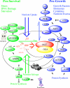
Figure 5. O-GlcNAc cycling and crosstalk with Multiple Cellular Nutrient-responsive pathways
The Hexosamine signaling pathway terminating in the enzymes of O-GlcNAc cycling lies at the center of many of the major nutrient sensing pathways (p38 MAPK, Insulin-AKT, mTOR, AMPK and SirT1). Broadly speaking these pathways can be considered either ‘Pro-survival’ cascades (shown in green), triggered by stress, DNA damage or starvation or ‘Pro-growth’ cascades (shown in blue) triggered by hormones, cytokines, growth factors, and nutrient excess. The pro-survival cascade limits protein synthesis and triggers nutrient salvage by proteasomal degradation and autophagy. In contrast, the pro-growth cascades enhance protein synthesis and block proteasome activation and autophagy. OGT is recruited to these two cascades by either interacting with PIP3 resulting from PI3K activation (blue double arrow) or by interacting with activated p38 MAPK (green double arrow). These mutually exclusive interactions of OGT produce a binary switch, bringing the nutrient sensing hexosamine signaling module to bear on the two major cascades as indicated in the text. For example, by blunting the action of AKT, OGT limits the pro-growth cascade. OGT also inhibits the proteasome. In contrast, binding to activated p38 MAPK, OGT changes its substrate specificity bringing its nutrient-sensing capabilities to bear on limited stress-specific targets (see text). The net result of these interactions is either nutrient uptake or nutrient salvage and sparing.
Figure 6
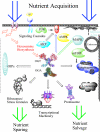
Figure 6. Key intracellular targets of O-GlcNAc impact nutrient uptake and salvage
As a modulator of signaling, endocytosis, transcription, translation and protein stability, the HSP may coordinately regulate nutrient sparing and salvage. Some of the key intracellular targets of O-GlcNAc modification are shown with respect to their role in nutrient uptake and salvage. OGT modifies numerous signaling cascades including those triggered by PI3K, MAPK, AMPK and mTOR (For details, see figure 5). In addition, by interacting with adapters like the D. melanogaster protein Milton, OGT may modulate intracellular movement of mitochondria on microtubules. The mammalian Milton homolog GRIF-1 binds to OGT and may be involved in orchestrating directed organelle movement including the trafficking of neurosecretory vesicles, endosomes, lysosomes and autophagosomes. These processes are critical for nutrient acquisition and immunity. OGT overexpression alters the movement of GLUT4 rich endosomes to the plasma membrane in response to insulin, a hallmark of mammalian insulin resistance. O-GlcNAc may also play a role in the switch from anabolic to catabolic metabolism. For example, O-GlcNAc modifies many ribosomal proteins and is required for the formation of stress granules involved in mRNA triage. O-GlcNAc also regulates the proteasome by modifying the ATP-dependent rpt2 domain as described in the text, and may directly, or indirectly regulate autophagy. O-GlcNAc also modifies nuclear pores and transcription-complexes playing an essential role in transcriptional repression [125].

References

    1. Love DC, Hanover JA. The hexosamine signaling pathway: deciphering the “O-GlcNAc code”. Sci STKE. 2005;2005:re13. - PubMed
    1. Hart GW, Housley MP, Slawson C. Cycling of O-linked beta-N-acetylglucosamine on nucleocytoplasmic proteins. Nature. 2007;446:1017–1022. - PubMed
    1. Roos MD, Hanover JA. Structure of O-linked GlcNAc transferase: mediator of glycan-dependent signaling. Biochem Biophys Res Commun. 2000;271:275–280. - PubMed
    1. Banerjee S, Robbins PW, Samuelson J. Molecular characterization of nucleocytosolic O-GlcNAc transferases of Giardia lamblia and Cryptosporidium parvum. Glycobiology. 2009;19:331–336. - PMC - PubMed
    1. Hanover JA. Glycan-dependent signaling: O-linked N-acetylglucosamine. FASEB J. 2001;15:1865–1876. - PubMed

MeSH terms

LinkOut - more resources