. 2009 Jul 31;35(2):143-53.
doi: 10.1016/j.molcel.2009.05.029.

A quorum-sensing antagonist targets both membrane-bound and cytoplasmic receptors and controls bacterial pathogenicity

Affiliations

A quorum-sensing antagonist targets both membrane-bound and cytoplasmic receptors and controls bacterial pathogenicity

Lee R Swem et al. Mol Cell. .

Abstract

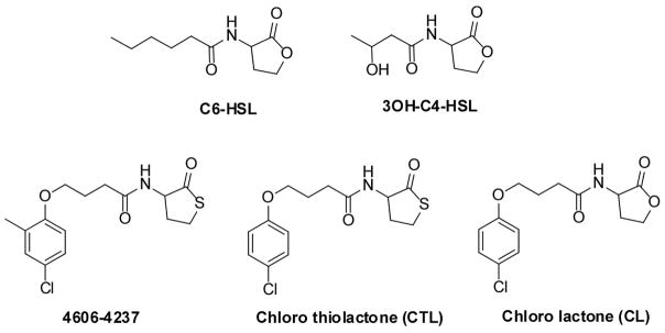
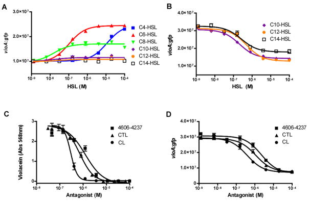
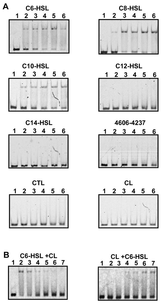
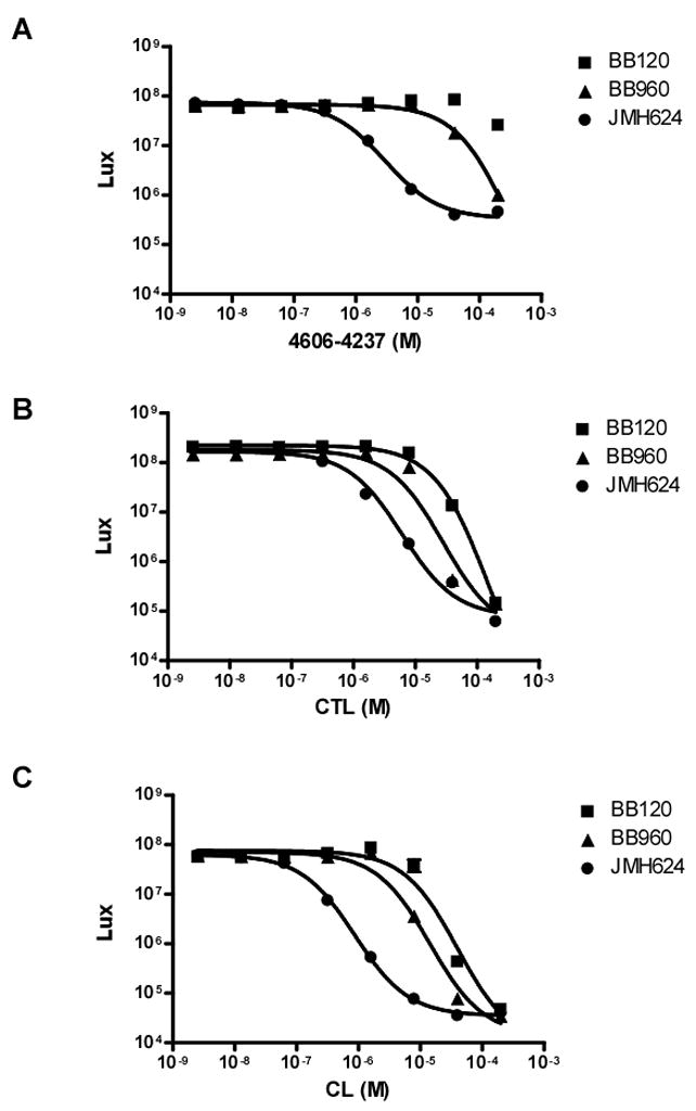
Quorum sensing is a process of bacterial communication involving production and detection of secreted molecules called autoinducers. Gram-negative bacteria use acyl-homoserine lactone (AHL) autoinducers, which are detected by one of two receptor types. First, cytoplasmic LuxR-type receptors bind accumulated intracellular AHLs. AHL-LuxR complexes bind DNA and alter gene expression. Second, membrane-bound LuxN-type receptors bind accumulated extracellular AHLs. AHL-LuxN complexes relay information internally by phosphorylation cascades that direct gene expression changes. Here, we show that a small molecule, previously identified as an antagonist of LuxN-type receptors, is also a potent antagonist of the LuxR family, despite differences in receptor structure, localization, AHL specificity, and signaling mechanism. Derivatives were synthesized and optimized for potency, and in each case, we characterized the mode of action of antagonism. The most potent antagonist protects Caenorhabditis elegans from quorum-sensing-mediated killing by Chromobacterium violaceum, validating the notion that targeting quorum sensing has potential for antimicrobial drug development.


Figures

Figure 1
Figure 1. The C. violaceum and V. harveyi Quorum-Sensing Circuits
Left panel: The cytoplasmic quorum-sensing receptor, CviR, from C. violaceum binds to the AHL autoinducer (black ovals) at high cell density (HCD). The CviR-AHL complex binds to DNA and activates expression of the vio genes required for production of the purple pigment, violacein. CviI is the C6-HSL synthase. Right panel: The membrane-bound quorum-sensing receptor, LuxN from V. harveyi binds to the AHL autoinducer (black ovals) at high cell density (HCD) resulting in a phosphorylation cascade that activates expression of the lux genes required for bioluminescence. LuxM is the 3OH-C4-HSL autoinducer synthase.
Figure 2
Figure 2. Structures of the Quorum-Sensing Autoinducers and Synthetic Antagonists
Structures and designations of the quorum-sensing autoinducers and synthetic antagonists for C. violaceum and V. harveyi.
Figure 3
Figure 3. CviR Dose-Response Curves
(A) CviR-dependent vioA-gfp expression in E. coli is plotted as a function of concentration of the specified homoserine lactone (HSL) molecules. (B) Inhibition of CviR-dependent vioA-gfp expression in E. coli is plotted as a function of the concentration of the specified molecule in the presence of a 500 nM C6-HSL. (C) CviR-dependent violacein production in wild type C. violaceum is plotted as a function of specified antagonist molecule. (D) Inhibition of CviR-dependent vioA-gfp expression in E. coli is plotted as a function of the specified antagonist molecule. In all panels, data were fit with a variable-slope sigmoidal dose-response curve to determine EC50 or IC50 values. Error bars represent the standard error of the mean for three independent trials.
Figure 4
Figure 4. Solubility Analysis of CviR Bound to Ligands
(A) SDS-PAGE analysis of E. coli whole cell (W) and soluble (S) extracts of cell cultures expressing CviR in the presence of dimethyl sulfoxide (DMSO) (Lanes 2 and 3), C6-HSL (Lanes 4 and 5), and C10-HSL (Lanes 6 and 7). (B) SDS-PAGE analysis of E. coli whole cell (W) and soluble (S) extracts of cell cultures expressing CviR in the presence of DMSO (Lanes 2 and 3), 4606-4237 (Lanes 4 and 5), Chloro-thiolactone (CTL) (Lanes 6 and 7) and Chlorolactone (CL) (Lanes 8 and 9). The L above the first lane designates the molecular weight ladder.
Figure 5
Figure 5. Gel Mobility Shift Analysis of the CviR Protein Bound to Agonist and Antagonist Molecules
(A) CviR binding to the vioA promoter at concentrations of 0 nM (Lane 1), 100 nM (Lane 2), 200 nM (Lane 3), 300 nM (Lane 4), 400 nM (Lane 5), and 500 nM (Lane 6). Each panel corresponds to CviR loaded with a different molecule. (B) CviR proteins loaded with C6-HSL and loaded with CL at concentrations of 500 nM were incubated for 20 min with 0, 0.5, 1, 3, 5 or 10 μM CL and C6-HSL, respectively. The vioA probe was added and allowed to incubate at room temperature for 20 additional min prior to being subjected to electrophoresis. No protein (Lane 1), 0 μM (Lane 2), 0.5 μM (Lane 3), 1 μM (Lane 4), 3 μM (Lane 5), 5 μM (Lane 6), 10 μM (Lane 7) CL or C6-HSL.
Figure 6
Figure 6. V. harveyi Bioluminescence in Response to Antagonists
Light production from wild type V. harveyi (BB120), a luxPQ mutant (BB960), and a luxPQ, luxM double mutant (JMH624) was measured in the presence of the specified concentrations of 4606-4237 (A), CTL (B), or (CL) (C). Data were fit with a variable-slope sigmoidal dose-response curve to determine IC50 values. Error bars although small are included and represent the standard error of the mean for three independent trials.
Figure 7
Figure 7. C. elegans Survival Following C. violaceum Infection and Treatment
(A) Kaplan-Meier survival curve of a C. elegans population infected with C. violaceum cviI mutant in the presence of the control solution of dimethyl sulfoxide (DMSO) or the specified molecules. (B) Kaplan-Meier survival curve of a C. elegans population infected with wild type C. violaceum in the absence of any quorum-sensing antagonist or in the presence of CL.

References

    1. Aballay A, Ausubel FM. Caenorhabditis elegans as a host for the study of host-pathogen interactions. Curr Opin Microbiol. 2002;5:97–101. - PubMed
    1. Bassler BL, Wright M, Showalter RE, Silverman MR. Intercellular signalling in Vibrio harveyi: sequence and function of genes regulating expression of luminescence. Mol Microbiol. 1993;9:773–786. - PubMed
    1. Bassler BL, Wright M, Silverman MR. Multiple signalling systems controlling expression of luminescence in Vibrio harveyi: sequence and function of genes encoding a second sensory pathway. Mol Microbiol. 1994;13:273–286. - PubMed
    1. Cao JG, Meighen EA. Purification and structural identification of an autoinducer for the luminescence system of Vibrio harveyi. J Biol Chem. 1989;264:21670–21676. - PubMed
    1. Dhakal BK, Lee W, Kim YR, Choy HE, Ahnn J, Rhee JH. Caenorhabditis elegans as a simple model host for Vibrio vulnificus infection. Biochem Biophys Res Commun. 2006;346:751–757. - PubMed

Publication types

MeSH terms