. 2009 Jul 31;35(2):164-80.
doi: 10.1016/j.molcel.2009.05.028.

Two-site phosphorylation of EPRS coordinates multimodal regulation of noncanonical translational control activity

Affiliations

Two-site phosphorylation of EPRS coordinates multimodal regulation of noncanonical translational control activity

Abul Arif et al. Mol Cell. .

Abstract

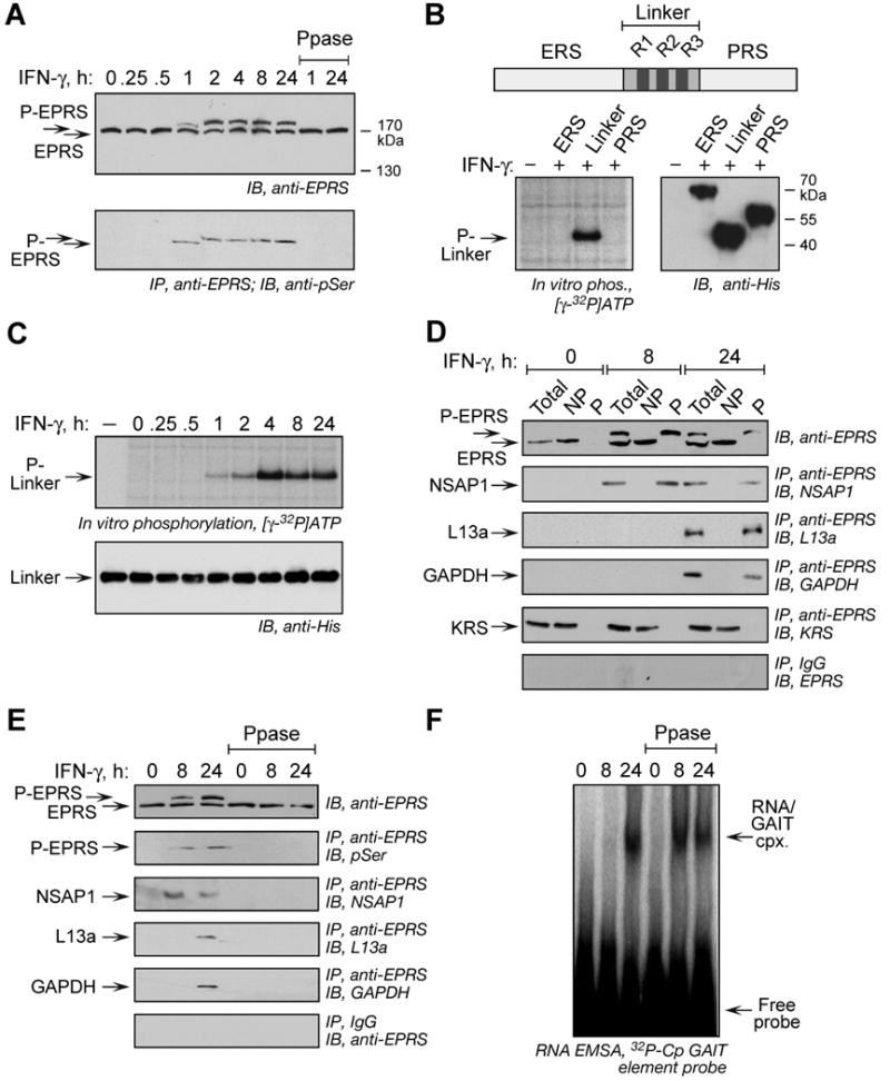
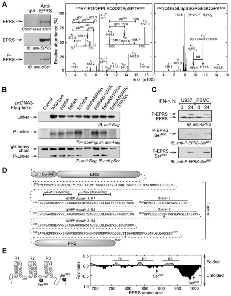
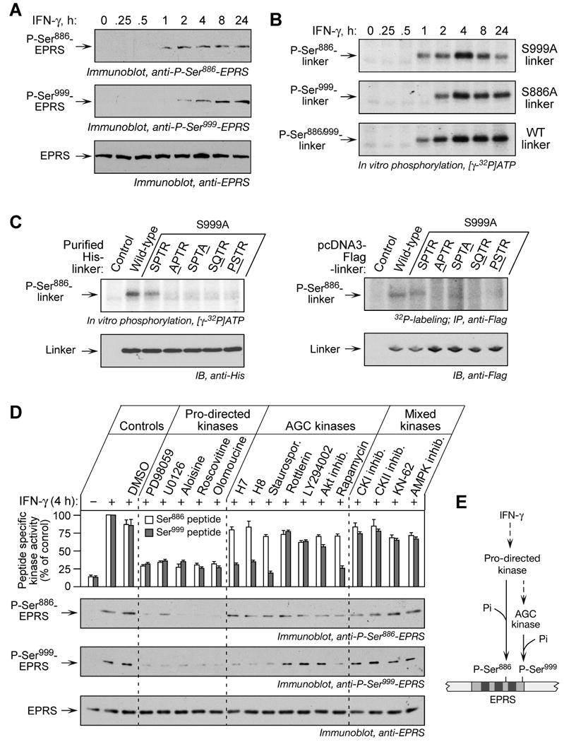
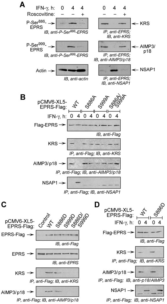
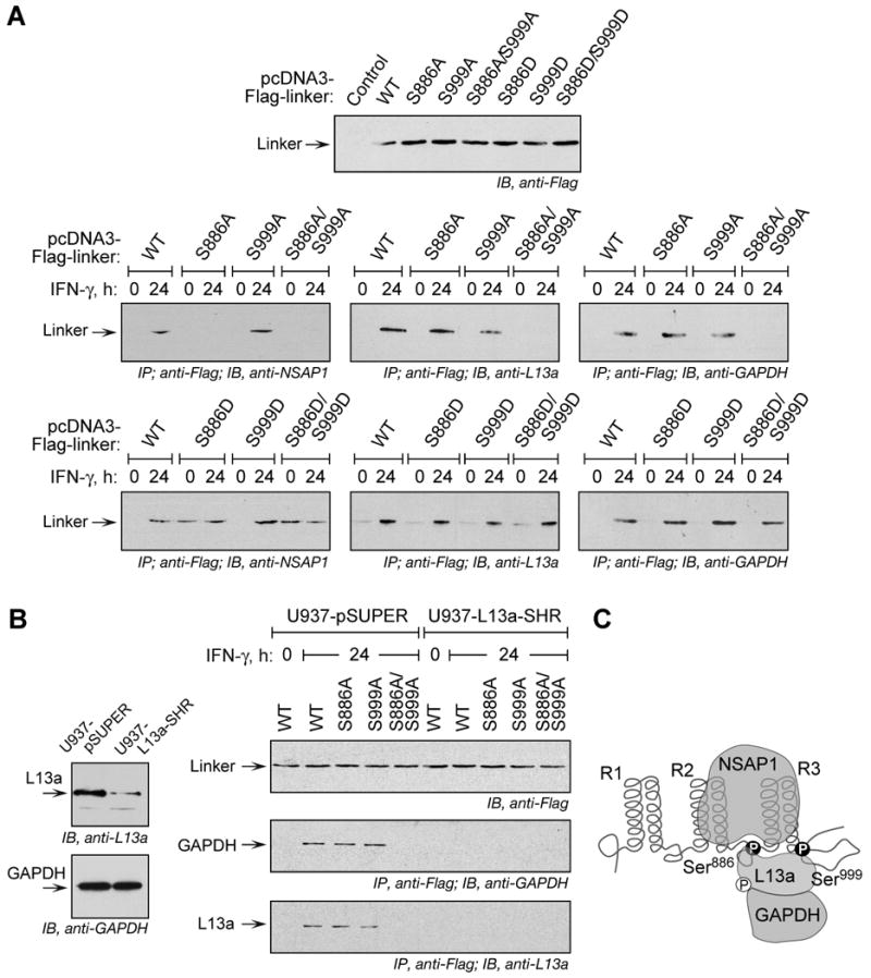
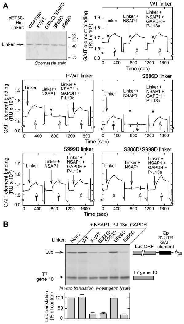
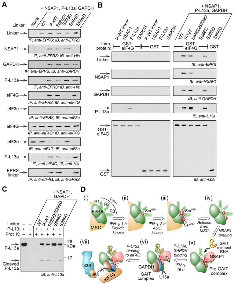
Glutamyl-prolyl tRNA synthetase (EPRS) is a component of the heterotetrameric gamma-interferon-activated inhibitor of translation (GAIT) complex that binds 3'UTR GAIT elements in multiple interferon-gamma (IFN-gamma)-inducible mRNAs and suppresses their translation. Here, we elucidate the specific EPRS phosphorylation events that regulate GAIT-mediated gene silencing. IFN-gamma induces sequential phosphorylation of Ser(886) and Ser(999) in the noncatalytic linker connecting the synthetase cores. Phosphorylation of both sites is essential for EPRS release from the parent tRNA multisynthetase complex. Ser(886) phosphorylation is required for the interaction of NSAP1, which blocks EPRS binding to target mRNAs. The same phosphorylation event induces subsequent binding of ribosomal protein L13a and GAPDH and restores mRNA binding. Finally, Ser(999) phosphorylation directs the formation of a functional GAIT complex that binds initiation factor eIF4G and represses translation. Thus, two-site phosphorylation provides structural and functional pliability to EPRS and choreographs the repertoire of activities that regulates inflammatory gene expression.


Figures

Figure 1
Figure 1. Inducible, Multisite Serine Phosphorylation in EPRS Linker Domain
(A) Multisite EPRS phosphorylation. Phosphorylated EPRS (P-EPRS) in lysates from IFN-γ-treated U937 cells was resolved by SDS-PAGE (4.5% polyacrylamide) and detected by immunoblot with anti-EPRS antibody. Two samples were incubated with shrimp alkaline phosphatase (PPase) before immunoblot analysis. The same lysates were immunoprecipitated with anti-EPRS antibody and probed with anti-pSer antibody. (B) The non-catalytic EPRS linker domain is phosphorylated. Schematic of major human EPRS domains. In vitro phosphorylation of His-tagged EPRS domains by lysates of 24-h, IFN-γ-treated U937 cells. All domains detected by immunoblot with anti-His antibody. (C) Lysates from IFN-γ treated cells were used to in vitro phosphorylate recombinant, His-tagged EPRS linker. Linker was detected by immunoblot with anti-His antibody. (D) EPRS phosphorylation is required for release from MSC and interaction with GAIT proteins. Lysates from IFN-γ treated U937 cells were applied to phosphoprotein affinity column to isolate P-EPRS and unmodified EPRS. Total, non-phosphorylated (NP), and phosphorylated (P) fractions were assessed by immunoblot with anti-EPRS antibody. The same fractions were immunoprecipitated with anti-EPRS and detected with antibodies against GAIT constituent proteins and the MSC constituent KRS. As a control, fractions were immunoprecipitated with rabbit IgG and immunoblotted with anti-EPRS. (E) Dephosphorylation of EPRS disrupts the pre-GAIT and GAIT complexes. Lysates from IFN-γ-treated U937 cells were incubated with shrimp alkaline phosphatase and dephosphorylation detected with anti-EPRS antibody, and by immunoprecipitation with anti-EPRS and immunoblot with anti-phosphoSer. Precipitates were probed with antibodies against all GAIT proteins. (F) EPRS phosphorylation is not required for binding to GAIT element. EPRS binding to GAIT element was determined by RNA EMSA using 32P-labeled Cp GAIT element RNA probe in control and phosphatase-treated lysates of IFN-γ-activated cells.
Figure 2
Figure 2. IFN-γ Induces Phosphorylation of Ser886 and Ser999 in EPRS Linker
(A) Mass spectrometric identification of phosphorylation sites. Phospho-EPRS was isolated from 24-h, IFN-γ-treated U937 cells by phosphoprotein affinity column and immunoprecipitation with anti-EPRS antibody. Phosphorylation was shown by immunoblot with anti-pSer antibody (left). Excised, Coomassie-stained P-EPRS was subjected to in-gel tryptic digest, and CID spectra obtained from LC-tandem MS identified two phospho-peptides. CID spectra of 871EYIPGQPPLSQSSDSSPTR889 were dominated by fragmentation at several Pro residues, including Pro874 forming the y16+2 ion and Pro877 to give y13 ion (center). Other sequence-specific y and b ions limited the phospho-site to Ser885 or Ser886. Detailed inspection indicated a low-abundance y4 (m/z 540) and b15 (m/z 1586) ions (insets). The latter indicated lack of phosphorylation at Ser885, and the 167 Da mass difference between y4 and y3 ions indicated Ser886 as the phosphorylation site. The fragmentation spectra of 992NQGGGLSSSGAGEGQGPK1009 were dominated by [M+2H]+2-H3PO4 ion at m/z 785.5 (right). Low-abundance, sequence-specific y-ions were on both sides of three potential phosphorylation sites in 998SSS1000. Ser999 was identified as the likely phosphorylation site by the y11 ion at m/z 956.4, which differs from y12 ion at m/z 1141.5 by 185 Da, consistent with the loss of unmodified Ser998 (87 Da) and H3PO4 (98 Da). Also, MS3 analysis of the ion at m/z 785.5 showed fragment ions indicating Ser999 as the dehydroalanine residue resulting from loss of H3PO4 of a phosphoserine residue (not shown). (B) Verification of phosphorylation sites. N-terminus-Flag-tagged, wild-type and mutant linkers in pcDNA3 vector were transiently transfected into U937 cells, and expression was determined by immunoblot. Cells were incubated for 4 h with 32P-orthophosphate and IFN-γ. Lysates were immunoprecipitated with anti-Flag antibody, and phosphorylation determined by autoradiography, and by detection with anti-pSer antibody. (C) Phospho-specific EPRS antibodies confirm inducible, 2-site phosphorylation. Phospho-specific antibodies raised against peptides containing pSer886 and pSer999 were used to probe lysates from IFN-γ-treated U937 cells and peripheral blood mononuclear cells (PBMC). (D) Sequence and domains of human EPRS linker. The N-terminus EF1Bγ-like domain and ERS domain are connected to the C-terminus PRS domain by a linker consisting of three WHEP repeats, R1, R2, and R3, separated by two spacers. Phosphorylation sites indicated by “*”. (E) Schematic of two phosphorylation sites in EPRS linker domain (left). Predicted folding probability of EPRS linker polypeptide sequence determined by FoldIndex using a window size of 51 (right).
Figure 3
Figure 3. Ordered Phosphorylation of Ser886 and Ser999 by two Ser/Thr kinases
(A) Ser886 phosphorylation precedes Ser999 phosphorylation. Lysates from U937 cells treated with IFN-γ were resolved by SDS-PAGE (12% polyacrylamide) and analyzed by immunoblot with phospho-specific antibodies, and with anti-EPRS antibody. (B) Ser886 and Ser999 phosphorylation events are independent. Lysates of IFN-γ-treated U937 cells were used to in vitro phosphorylate His-tagged wild-type and mutant linkers. (C) Identification of the kinase recognition motif for Ser886 phosphorylation. Lysates of 4-h, IFN-γ-treated cells were used to in vitro phosphorylate purified His-tagged wild-type and linkers with mutations in key motif amino acids; equal linker amount was detected with anti-His antibody (left panels). The pcDNA3-Flag-linker DNAs containing motif mutations were transiently expressed in U937 cells and labeled with 32P-orthophosphate in presence of IFN-γ for 4 h. Lysates were immunoprecipitated with anti-Flag antibody, and 32P incorporation determined by autoradiography (right panels). (D) Distinct kinases phosphorylate Ser886 and Ser999. U937 cells were incubated with IFN-γ for 0.5 h and then with kinase inhibitors for an additional 3.5 h. Cells incubated with or without IFN-γ and dimethyl sulfoxide (DMSO, 0.5%) served as controls. Pro-directed kinase group inhibitors were PD98059 (10 μM), U0126 (10 μM), aloisine (5 μM), roscovitine (10 μM), and olomoucine (50 μM). AGC kinase group inhibitors were H-7 (10 μM), H-8 (10 μM), staurosporine (Staurospor. 20 nM), rottlerin (100 μM), LY294002 (2.5 μM), Akt inhibitor-VIII (2.5 μM), rapamycin (10 nM). Other inhibitors were CKI inhibitor (100 μM), CKII inhibitor (100 μM), KN-62 (5 μM), and AMPK inhibitor (2 μM). Lysate kinase activity was determined as phosphorylation of Ser886 (white) and Ser999 (grey) phospho-acceptor peptides, and expressed as % of control lysates (mean ± SEM, top panel). Lysates were resolved on SDS-PAGE (12% polyacrylamide) and analyzed by immunoblot with indicated antibodies (panels 2-4). (E) Schematic of kinase pathway phosphorylating EPRS Ser886 and Ser999.
Figure 4
Figure 4. EPRS Phosphorylation is Required for Release from MSC
(A) Inhibition of phosphorylation blocks EPRS release from MSC. U937 cells were treated with IFN-γ and roscovitine or DSMO as in Fig. 3D. Lysates were immunoblotted with antibodies indicated. Same lysates were immunoprecipitated with anti-EPRS antibody and immunoblotted with antibodies against MSC components, KRS and AIMP3/p18, or against the pre-GAIT complex protein, NSAP1. (B) Both Ser phosphorylation events are required for EPRS release from MSC. Flag-tagged, full-length, wild-type (WT) or phosphorylation-defective (Ser-to-Ala, S-A) EPRS mutants were cloned into pCMV6-XL5 vector and expressed in U937 cells by transient transfection. After 18 h, cycloheximide (50 μg/ml, 30 min) was added to block new synthesis, and then incubated with IFN-γ for 4 h. Lysates were immunoprecipitated with anti-Flag antibody and immunoblotted with antibodies indicated. (C) Incorporation of EPRS phospho-mimetics into MSC. Flag-tagged, full-length, WT and phospho-mimetic (Ser-to-Asp, S-D) mutant EPRS was expressed in U937 cells by transient transfection as in (B) and measured by immunoblot with anti-Flag antibody. Endogenous EPRS was detected by anti-EPRS antibody. Lysates were immunoprecipitated with anti-Flag antibody and immunoblotted with antibodies indicated. (D) Release of S886D phospho-mimetic establishes dual-site phosphorylation-dependent release of EPRS from MSC. Cells expressing Flag-tagged, full-length EPRS were incubated with IFN-γ for 4 h. Lysates were immunoprecipitated with anti-Flag antibody and immunoblotted with antibodies indicated.
Figure 5
Figure 5. Role of Specific EPRS-Ser Phosphorylation in GAIT Complex Assembly
(A) Phosphorylation of Ser886 is essential for EPRS interaction with NSAP1, and phosphorylation of either Ser886 or Ser999 is essential for EPRS interaction with L13a and GAPDH. Flag-tagged wild type (WT), phospho-defective (Ser-to-Ala, S-A) and phospho-mimetic (Ser-to-Asp, S-D) mutant linkers were expressed by transient transfection and determined by immunoblot with anti-Flag antibody. After IFN-γ treatment, lysates were immunoprecipitated with anti-Flag antibody and immunoblotted with antibodies against NSAP-1, L13a, and GAPDH. (B) GAPDH interaction with phospho-EPRS requires L13a. Immunoblot showing depletion of L13a, and unaltered expression of GAPDH, in U937 cells with stable knockdown of L13a (U937-L13a-SHR) and vector control cells (U937-pSUPER) (left). Cells were transiently transfected with Flag-tagged WT and phosphorylation-defective EPRS linker constructs (right). Expression was determined by immunoblot with anti-Flag antibody. The cells were incubated with IFN-γ and lysates immunoprecipitated with anti-Flag antibody followed by immunoblot with anti-GAPDH or anti-L13a antibodies. (C) Schematic depicting interaction of NSAP1, L13a, and GAPDH to EPRS linker phosphorylated at Ser886 and Ser999.
Figure 6
Figure 6. Role of EPRS Linker Phosphorylation in Binding the GAIT RNA Element and GAIT-mediated Translational Silencing
(A) Ser886 phosphorylation is sufficient for reconstituting binding to GAIT RNA element. N-terminus, His-tagged wild-type (WT) and phospho-mimetic (Ser-to-Asp, S-D) mutant linkers were expressed in E. coli and purified by Ni-affinity chromatography. Phosphorylated, wild-type (P-WT) linker was obtained by in vitro phosphorylation of purified protein with lysates of 4-h, IFN-γ-treated U937 cells, and repurification by Ni-affinity column. Biotinylated Cp GAIT RNA element was immobilized on a streptavidin sensor chip, and binding of linker to RNA was determined by SPR and expressed as resonance units (RU). After removal of bound protein with wash buffer (open arrows), binding of linkers preincubated with NSAP1 was measured, then linkers preincubated with NSAP1, GAPDH, and phospho-L13a (P-L13a) (each at 10-fold molar excess). (B) Ser999 phosphorylation is critical for GAIT complex activation. His-tagged, wild-type (WT), phosphorylated-WT (P-WT), and phospho-mimetic linker proteins were preincubated with His-tagged NSAP1, GAPDH, and phosphorylated, His-tagged L13a (P-L13a), and then added to wheat germ extract for in vitro translation of luciferase (Luc)-Cp-GAIT element reporter in presence of 35S-labeled Met. T7 gene 10 RNA was co-translated as a specificity control. Luc expression was quantified by densitometry, normalized by T7 gene 10 translation, and expressed as % of control (mean ± SEM, n = 3 experiments).
Figure 7
Figure 7. Ser999 Phosphorylation is Required for L13a-mediated Interaction of GAIT Complex with eIF4G
(A) Ser999 phosphorylation is required for GAIT complex interaction with eIF4G during in vitro translation. In vitro translation of GAIT element-bearing reporter in wheat germ extract as in Figure 7B. The reaction mixture was immunoprecipitated with anti-EPRS and eIF4G antibodies after digestion with RNase A/T1, and immunoblotted with antibodies indicated. (B) Ser999 phosphorylation facilitates P-L13a-mediated interaction of GAIT complex with recombinant eIF4G. Individual (left panels) or combined (right panels) GAIT complex proteins were incubated with GST-eIF4G or GST immobilized to glutathione-agarose beads. After washing, the interaction was analyzed by immunoblotting with antibodies against GAIT components and eIF4G. (C) EPRS Ser999 phosphorylation alters protease susceptibility of L13a. P-L13a (150 ng) was preincubated with GAIT components including mutant EPRS linkers and then treated with 10 ng proteinase (Prot.) K for 15 min at 30°C. Untreated and protease-treated P-L13a served as controls. Cleavage was determined by immunoblot with anti-L13a antibody. (D) Schematic illustrating function of 2-site phosphorylation of EPRS (i) EPRS resides in the MSC. (ii) IFN-γ induces phosphorylation of Ser886 by a proline-directed kinase. (iii) IFN-γ induces phosphorylation of Ser999 by an AGC kinase group member. Phosphorylation of both Ser886 and Ser999 (highlighted with radial lines) induces EPRS release from the MSC. (iv) Diphosphorylated EPRS translocates from the MSC to the cytoplasm. (v) Phosphorylation of EPRS Ser886 is required for NSAP1 binding that blocks binding of EPRS to the GAIT RNA element. (vi) EPRS Ser886 phosphorylation is required for binding of phospho-L13a and GAPDH that induces a conformational shift and permits GAIT RNA element binding to EPRS. (vii) Phosphorylation of EPRS Ser999 is required for translational silencing activity of GAIT complex by permitting interaction of phospho-L13a with eIF4G, and blocking the binding of eIF3 of the 43S ribosomal complex to eIF4G.

Similar articles

Cited by

References

    1. Amanchy R, Periaswamy B, Mathivanan S, Reddy R, Tattikota SG, Pandey A. A curated compendium of phosphorylation motifs. Nat Biotechnol. 2007;25:285–286. - PubMed
    1. Beausoleil SA, Jedrychowski M, Schwartz D, Elias JE, Villen J, Li J, Cohn MA, Cantley LC, Gygi SP. Large-scale characterization of HeLa cell nuclear phosphoproteins. Proc Natl Acad Sci U S A. 2004;101:12130–12135. - PMC - PubMed
    1. Berg BH. The early influence of 17-β-oestradiol on 17 aminoacyl-tRNA synthetases of mouse uterus and liver. Phosphorylation as a regulation mechanism. Biochim Biophys Acta. 1977;479:152–171. - PubMed
    1. Cahuzac B, Berthonneau E, Birlirakis N, Guittet E, Mirande M. A recurrent RNA-binding domain is appended to eukaryotic aminoacyl-tRNA synthetases. EMBO J. 2000;19:445–452. - PMC - PubMed
    1. Casnellie JE. Assay of protein kinases using peptides with basic residues for phosphocellulose binding. Methods Enzymol. 1991;200:115–120. - PubMed

Publication types

MeSH terms