

The effect of significant tumor reduction on the dose distribution in intensity modulated radiation therapy for head-and-neck cancer: a case study
- PMID: 19647637
- PMCID: PMC2892776
- DOI: 10.1016/j.meddos.2008.10.004
The effect of significant tumor reduction on the dose distribution in intensity modulated radiation therapy for head-and-neck cancer: a case study
Abstract
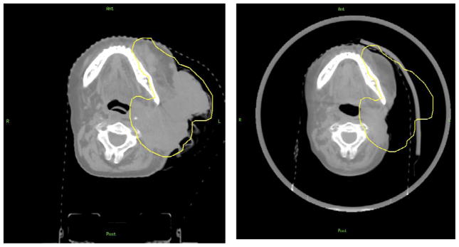
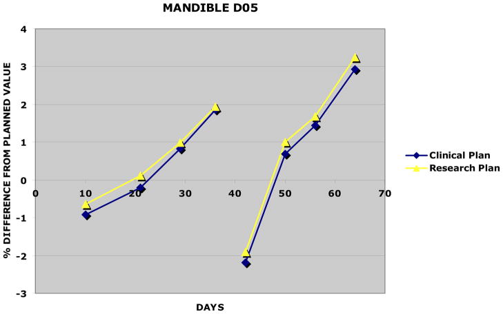
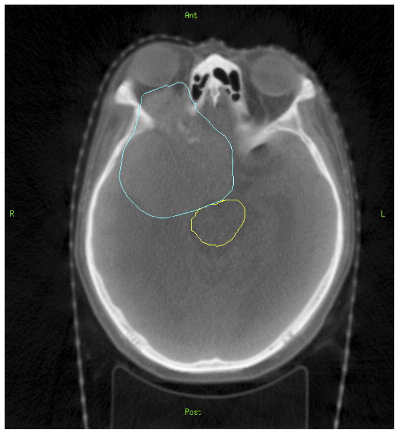
We present a unique case in which a patient with significant tissue loss was monitored for dosimetric changes using weekly cone beam computed tomography (CBCT) scans. A previously treated nasopharynx patient presented with a large, exophytic, recurrent left neck mass. The patient underwent re-irradiation to 70 Gy using intensity modulated radiation therapy (IMRT) with shielding blocks over the spinal cord and brain stem. Weekly CBCT scans were acquired during treatment. Target contours and treatment fields were then transferred from the original treatment planning computed tomography (CT) to the CBCT scans and dose calculations were performed on all CBCT scans and compared to the planning doses. In addition, a "research" treatment plan was created that assumed the patient had not been previously treated, and the above analysis was repeated. Finally, to remove the effects of setup error, the outer contours of 2 CBCT scans with significant tumor reductions were transferred to the planning scan and dose in the planning scan was recalculated. Planning treatment volume (PTV) decreased 45% during treatment. Spinal cord D05 differed from the planned value by 3.5 +/- 9.8% (average + standard deviation). Mean dose to the oral cavity and D05 of the mandible differed from the planned value by 0.9 +/- 2.1% and 0.6 +/- 1.5%, respectively. Results for the research plan were comparable. Target coverage did not change appreciably (-0.2 +/- 2.5%). When the planning scan was recalculated with the reduced outer contour from the CBCT, spinal cord D05 decreased slightly due to the reduction in scattered dose. Weekly imaging provided us the unique opportunity to use different methods to examine the dosimetric effects of an unusually large loss of tissue. We did not see that tissue loss alone resulted in a significant effect on the dose delivered to the spinal cord for this case, as most fluctuation was due to setup error. In the IGRT era, delivered dose distributions can be more readily determined during treatment, and this information can be useful in deciding whether replanning is necessary.
Figures


References
-
- Chui C, Spirou SV. Inverse planning algorithms for external beam radiation therapy. Med Dosim. 2001;26:189–97. - PubMed
-
- Eisbruch A. Intensity-modulated radiation therapy: A clinical perspective. Semin Radiat Oncol. 2002;12:197–8. - PubMed
-
- Nutting C, Dearnaley DP, Webb S. Intensity modulated radio-therapy: A clinical review. Br J Radiol. 2000;73:465–9. - PubMed
-
- Mechalakos J, Hunt M, Hong L, et al. Measurement of IMRT head and neck setup error using an on-board kilovotage imager. Int J Radiat Oncol Biol Phys. 2005;63:S353–4.
-
- Barker JL, Garden AS, Ang KK, et al. Quantification of volumetric and geometric changes occurring during fractionated radiotherapy for head-and-neck cancer using an integrated CT/linear accelerator system. Int J Radiat Oncol Biol Phys. 2004;59:960–70. - PubMed
Publication types
MeSH terms
Grants and funding
LinkOut - more resources
Full Text Sources
