. 2009 Sep 25;284(39):26789-96.
doi: 10.1074/jbc.M109.026823. Epub 2009 Jul 31.

Inflammasome-dependent caspase-1 activation in cervical epithelial cells stimulates growth of the intracellular pathogen Chlamydia trachomatis

Affiliations

Inflammasome-dependent caspase-1 activation in cervical epithelial cells stimulates growth of the intracellular pathogen Chlamydia trachomatis

Ali A Abdul-Sater et al. J Biol Chem. .

Abstract

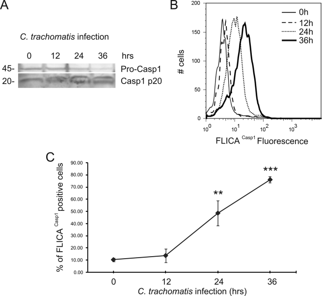
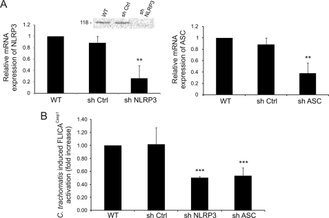
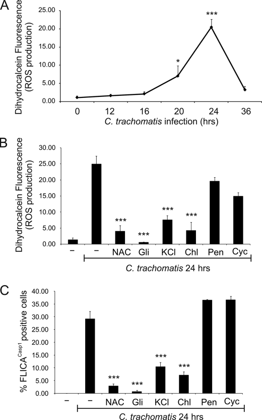
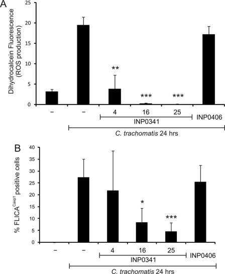
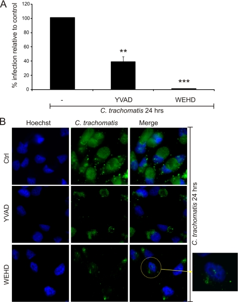
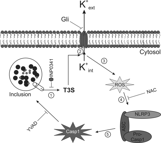
Inflammasomes have been extensively characterized in monocytes and macrophages, but not in epithelial cells, which are the preferred host cells for many pathogens. Here we show that cervical epithelial cells express a functional inflammasome. Infection of the cells by Chlamydia trachomatis leads to activation of caspase-1, through a process requiring the NOD-like receptor family member NLRP3 and the inflammasome adaptor protein ASC. Secretion of newly synthesized virulence proteins from the chlamydial vacuole through a type III secretion apparatus results in efflux of K(+) through glibenclamide-sensitive K(+) channels, which in turn stimulates production of reactive oxygen species. Elevated levels of reactive oxygen species are responsible for NLRP3-dependent caspase-1 activation in the infected cells. In monocytes and macrophages, caspase-1 is involved in processing and secretion of pro-inflammatory cytokines such as interleukin-1beta. However, in epithelial cells, which are not known to secrete large quantities of interleukin-1beta, caspase-1 has been shown previously to enhance lipid metabolism. Here we show that, in cervical epithelial cells, caspase-1 activation is required for optimal growth of the intracellular chlamydiae.


Figures

FIGURE 1.
FIGURE 1.
C. trachomatis induces caspase-1 activation in HeLa cells. HeLa cells were infected with C. trachomatis at an m.o.i. = 3 for 12, 24, and 36 h. A, Western blot analysis of HeLa cell lysates was performed to monitor caspase-1 (Casp1) activation using an antibody that detects pro-caspase-1 (p45, upper band) and active caspase-1 (p20, lower band). B, caspase-1 activation was quantified using fluorescent FLICACasp1 reagent and analyzed by flow cytometry. Nonfluorescent cells were gated in the first log-decade, and the fluorescence intensity was proportional to the level of caspase-1 activation. C, column chart of FLICACasp1 flow cytometry data, showing the % of cells with activated caspase-1 as a function of infection time. Error bars represent standard deviation (n = 3). ** indicate p < 0.01; *** indicate p < 0.001, compared with uninfected cells.
FIGURE 2.
FIGURE 2.
C. trachomatis-induced caspase-1 activation requires NLRP3 inflammasome. A, HeLa cells were stably transfected with shRNAs that target NLRP3 or ASC, and mRNA expressions of NLRP3 (left panel) and ASC (right panel) were quantified by real time PCR and compared with wild type (WT) and nontarget control (sh Ctrl). Inset, Western blot analysis of wild type HeLa cells, cells treated with nontarget control, and cells treated with shNLRP3, confirming decreased expression of the NLRP3 protein after mRNA knockdown. Western blot was performed with an anti-NLRP3 antibody, which detects the 118-kDa protein. B, NLRP3, ASC, or nontarget control knockdown HeLa cells were infected with L2 at an m.o.i. = 3 for 24 h, and C. trachomatis-induced caspase-1 activation was measured by FLICACasp1. The fold increase in caspase-1 activation in infected nontarget controls, shNLRP3-treated cells, and shASC-treated cells with respect to uninfected cells was compared with the increase in 24 h-infected wild type cells. Error bars represent standard deviation of an experiment performed in triplicate on three separate occasions. ** indicates p < 0.01; *** indicates p < 0.001.
FIGURE 3.
FIGURE 3.
Caspase-1 activation following C. trachomatis infection is caused by K+ efflux and ROS production. A, HeLa cells were infected with C. trachomatis at an m.o.i. = 3 for 0, 12, 16, 20, and 24 h, and intracellular ROS levels were measured with the fluorescent dihydrocalcein reagent and analyzed by flow cytometry. Data are plotted as a line chart. B and C, HeLa cells were infected or not with C. trachomatis at an m.o.i. = 3 for 24 h, and treated or not with 10 mm NAC, 50 μm glibenclamide (Gli), 70 mm KCl during the last 15 h of infection, or 60 μg/ml chloramphenicol (Chl), 100 μg/ml penicillin (Pen), 10 μg/ml cycloheximide (Cyc) during the last 6 h of infection. ROS production was quantified with dihydrocalcein (B), or caspase-1 activation was measured with FLICACasp1 (C). Error bars represent standard deviation from at least three separate experiments. * indicates p < 0.05; *** indicates p < 0.001, compared with uninfected cells (A) or untreated infected cells (B and C).
FIGURE 4.
FIGURE 4.
Chlamydial T3S is responsible for ROS production and caspase-1 activation. HeLa cells were infected or not with C. trachomatis at an m.o.i. = 3 for 24 h and treated or not with 4, 16, or 25 μm T3S inhibitor (INP0341) or 25 μm control INP (INP0406) for 9 h. ROS production was quantified by staining cells with dihydrocalcein (A), or caspase-1 activation was measured by staining cells with FLICACasp1 (B). Error bars represent standard deviation of at least three separate experiments. * indicates p < 0.05; ** indicates p < 0.01; *** indicates p < 0.001, compared with infected untreated cells.
FIGURE 5.
FIGURE 5.
Caspase-1 activation is required for efficient C. trachomatis infection. HeLa cells were infected with C. trachomatis at an m.o.i. = 3 for 24 h and treated or not with two different caspase inhibitors as follows: 100 μm caspase-1 inhibitor (Z-YVAD-fmk) or 100 μm caspase-1/caspase-5 inhibitor (Z-WEHD-fmk) at 9 h post infection. A, cells were harvested and retitrated on a fresh monolayer of HeLa cells for 24 h and stained with Hoechst stain for DNA and anti-chlamydial antibody, and infected cells were counted. B, cells were stained with Hoechst stain (blue) and anti-chlamydial antibody (green) and visualized on a fluorescence microscope. Ctrl, control. Error bars represent standard deviation of three separate experiments. ** indicates p < 0.01; *** indicates p < 0.001, compared with infected untreated cells.
FIGURE 6.
FIGURE 6.
C. trachomatis triggers inflammasome-mediated caspase-1 activation through T3S-induced K+ efflux and ROS production. Following infection, chlamydiae inject virulence factors via the T3S apparatus into the host cell cytosol, causing loss of intracellular potassium and resulting in the production of ROS. Elevated ROS levels trigger the assembly of the NLRP3 inflammasome, which subsequently activates caspase-1. Activated caspase-1 plays a role in lipid metabolism and glycolysis and enhances development of the chlamydiae.

References

    1. Gerbase A. C., Rowley J. T., Mertens T. E. (1998) Lancet 351, 2–4 - PubMed
    1. Miller W. C., Ford C. A., Morris M., Handcock M. S., Schmitz J. L., Hobbs M. M., Cohen M. S., Harris K. M., Udry J. R. (2004) JAMA 291, 2229–2236 - PubMed
    1. Schachter J. (1999) in Chlamydia: Intracellular Biology, Pathogenesis, and Immunity ( Stephens R. S. ed) pp. 139–170, American Society for Microbiology, Washington, DC
    1. Thylefors B., Négrel A. D., Pararajasegaram R., Dadzie K. Y. (1995) Bull. World Health Organ. 73, 115–121 - PMC - PubMed
    1. Belland R., Ojcius D. M., Byrne G. I. (2004) Nat. Rev. Microbiol. 2, 530–531 - PubMed

Publication types

MeSH terms

Substances