. 2010 Apr;20(4):859-72.
doi: 10.1093/cercor/bhp150. Epub 2009 Jul 31.

Mechanisms of working memory disruption by external interference

Affiliations

Mechanisms of working memory disruption by external interference

Wesley C Clapp et al. Cereb Cortex. 2010 Apr.

Abstract

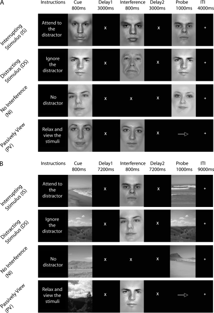
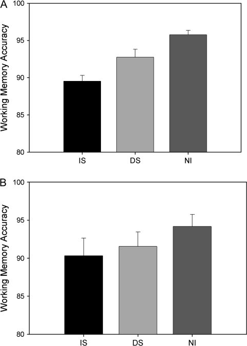
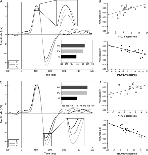
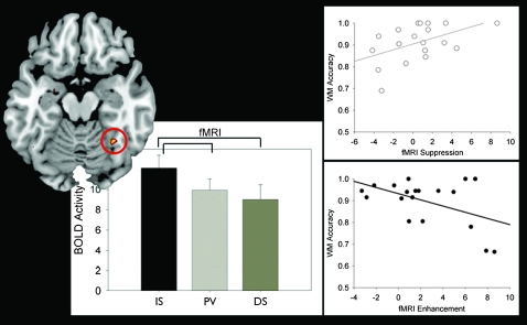
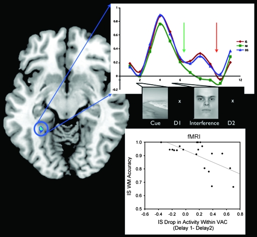
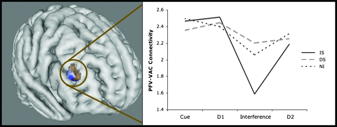
The negative impact of external interference on working memory (WM) performance is well documented; yet, the mechanisms underlying this disruption are not sufficiently understood. In this study, electroencephalogram and functional magnetic resonance imaging (fMRI) data were recorded in separate experiments that each introduced different types of visual interference during a period of WM maintenance: distraction (irrelevant stimuli) and interruption (stimuli that required attention). The data converged to reveal that regardless of the type of interference, the magnitude of processing interfering stimuli in the visual cortex (as rapidly as 100 ms) predicted subsequent WM recognition accuracy for stored items. fMRI connectivity analyses suggested that in the presence of distraction, encoded items were maintained throughout the delay period via connectivity between the middle frontal gyrus and visual association cortex, whereas memoranda were not maintained when subjects were interrupted but rather reactivated in the postinterruption period. These results elucidate the mechanisms of external interference on WM performance and highlight similarities and differences of distraction and multitasking.


Figures

Figure 1.
Figure 1.
Experimental paradigm. All participants performed 4 tasks, which were blocked and counterbalanced. (A) Experiment 1, EEG. (B) Experiment 2, fMRI.
Figure 2.
Figure 2.
Behavioral performance. (A) Experiment 1: WM accuracy. Participants performed best in the NI task, followed by DS, and then IS (all comparisons are significantly different, P < 0.05). (B) Experiment 2: WM accuracy. Accuracy was not significantly different in any of the tasks, but NI trended toward higher accuracy compared with IS (P < 0.1) and DS (P < 0.1) tasks.
Figure 3.
Figure 3.
Modulation of occipitotemporal EOI ERPs: (A and C) ERPs to interruptors (IS), passively viewed stimuli (PV), and distractors (DS). (A) P100 latency reveals significant enhancement. (B) The amount that participants allocate attention toward an interruptor (IS, enhancement) negatively correlates with their WM performance (R = −0.7, P < 0.001). Likewise, the amount of attention allocated away from a distractor (DS, suppression) positively correlates with WM (R = 0.5, P < 0.05). (C) N170 results showing significant enhancement of the N170 latency. (D) The same significant correlations were obtained as for the P100, such that the amount of attention allocated toward the interruptor and away from distractors predicts WM performance (R = −0.76, P < 0.0001; R = 0.64, P < 0.005, respectively). These results are replicated in the fMRI findings (Fig. 4).
Figure 4.
Figure 4.
FFA modulation and correlations with WM accuracy: The BOLD response in the FFA to interruptors (IS), passively viewed stimuli (PV), and distractors (DS) are presented in the bar graphs. The BOLD response was highest in response to the interruptors and lowest to the distractors (enhancement [IS > PV, P < 0.01]). Right panels: The amount that participants allocate attention toward an interruptor or away from a distractor (vs. passively viewed intervening stimuli) correlates with their WM performance (R = −0.54, P < 0.05; R = 0.53, P < 0.05, respectively). These results replicate the EEG findings (Fig. 3).
Figure 5.
Figure 5.
Time course of BOLD activity within the PPA and correlation with WM accuracy. The time series of the percent signal change in the PPA in IS, DS, and NI are plotted. The amount of decrement in the PPA signal in IS compared with DS is significant during delay2 (orange arrow, P < 0.05). Bottom right: the amount that PPA activity drops in IS between delay1 (green arrow) and delay2 negatively correlates with WM accuracy on the IS task.
Figure 6.
Figure 6.
MFG connectivity with PPA during WM maintenance. An area in the right MFG (brown) was identified with connectivity analysis using a PPA seed during the encoding period of the 3 WM tasks contrasted against PV (IS + DS + NI − 3PV). Connectivity between the PPA and the MFG area is maintained throughout the trial in both NI and DS whereas in IS connectivity declines during the interruption. A whole-brain correlation analysis using suppression indices as a regressor shows that in DS, stronger connectivity between the PPA and the right MFG (blue) is associated with greater suppression of the distractor. Right MFG is an ROI, not a statistical map, and the correlation analysis regressed with suppression has the cortex masked to highlight the area of interest.
Figure 7.
Figure 7.
Left IFG connectivity with FFA during interruption. An area in the left IFG (blue) was more active in IS than DS during the interference period with univariate analysis. Connectivity between this region (red) and the FFA was also found to correlate with enhancement of the BOLD signal in the FFA during interruption in the IS task. Cortical activity masked to highlight area of interest.

References

    1. Aron AR, Robbins TW, Poldrack RA. Inhibition and the right inferior frontal cortex. Trends Cogn Sci. 2004;8:170–177. - PubMed
    1. Baddeley A. Working memory. Oxford: Oxford University Press; 1986.
    1. Benjamini Y, Hochberg Y. Controlling the false discovery rate—a practical and powerful approach to multiple testing. J R Stat Soc Ser B. 1995;57:289–300.
    1. Bentin S, Allison T, Puce A, Perez E, McCarthy G. Electrophysiological studies of face perception in humans. J Cogn Neurosci. 1996;8:551–565. - PMC - PubMed
    1. Chao LL, Knight RT. Contribution of human prefrontal cortex to delay performance. J Cogn Neurosci. 1998;10:167–177. - PubMed

Publication types

MeSH terms