. 2009 Nov;297(5):L805-12.
doi: 10.1152/ajplung.00009.2009. Epub 2009 Jul 31.

N-acetylcysteine inhibits alveolar epithelial-mesenchymal transition

Affiliations

N-acetylcysteine inhibits alveolar epithelial-mesenchymal transition

V M Felton et al. Am J Physiol Lung Cell Mol Physiol. 2009 Nov.

Abstract

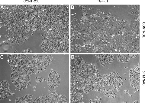
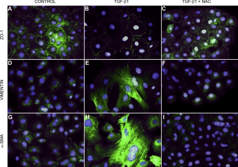
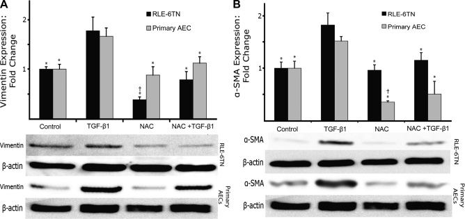
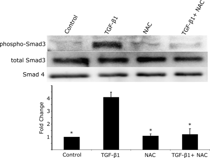
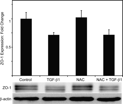
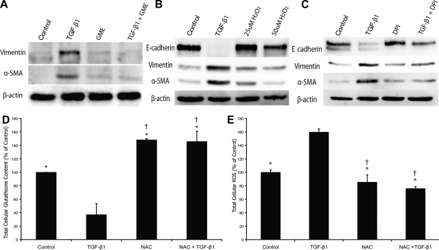
The ability of transforming growth factor-beta1 (TGF-beta1) to induce epithelial-mesenchymal transition (EMT) in alveolar epithelial cells (AEC) in vitro and in vivo, together with the demonstration of EMT in biopsies of idiopathic pulmonary fibrosis (IPF) patients, suggests a role for TGF-beta1-induced EMT in disease pathogenesis. We investigated the effects of N-acetylcysteine (NAC) on TGF-beta1-induced EMT in a rat epithelial cell line (RLE-6TN) and in primary rat alveolar epithelial cells (AEC). RLE-6TN cells exposed to TGF-beta1 for 5 days underwent EMT as evidenced by acquisition of a fibroblast-like morphology, downregulation of the epithelial-specific protein zonula occludens-1, and induction of the mesenchymal-specific proteins alpha-smooth muscle actin (alpha-SMA) and vimentin. These changes were inhibited by NAC, which also prevented Smad3 phosphorylation. Similarly, primary alveolar epithelial type II cells exposed to TGF-beta1 also underwent EMT that was prevented by NAC. TGF-beta1 decreased cellular GSH levels by 50-80%, whereas NAC restored them to approximately 150% of those found in TGF-beta1-treated cells. Treatment with glutathione monoethyl ester similarly prevented an increase in mesenchymal marker expression. Consistent with its role as an antioxidant and cellular redox stabilizer, NAC dramatically reduced intracellular reactive oxygen species production in the presence of TGF-beta1. Finally, inhibition of intracellular ROS generation during TGF-beta1 treatment prevented alveolar EMT, but treatment with H2O2 alone did not induce EMT. We conclude that NAC prevents EMT in AEC in vitro, at least in part through replenishment of intracellular GSH stores and limitation of TGF-beta1-induced intracellular ROS generation. We speculate that beneficial effects of NAC on pulmonary function in IPF may be mediated by inhibitory effects on alveolar EMT.


Figures

Fig. 1.
Fig. 1.
N-acetylcysteine (NAC) prevents transforming growth factor-β1 (TGF-β1)-induced changes in epithelial morphology in an alveolar epithelial cell (AEC) line (RLE-6TN). A: control cells in media alone demonstrate a cobblestone appearance and good cell-cell contacts consistent with an epithelial morphology. B: cells treated with TGF-β1 exhibit a fibroblast-like morphology with cellular elongation and reduction of cell-cell contacts. C: treatment with NAC alone causes no appreciable changes in cell morphology. D: NAC prevents changes induced by TGF-β1 and preserves an epithelial morphology.
Fig. 2.
Fig. 2.
NAC prevents aspects of alveolar epithelial-mesenchymal transition (EMT) in RLE-6TN cells. Control cells express significant amounts of membrane-associated ZO-1, but little vimentin and α-smooth muscle actin (α-SMA) (A, D, G). Cells treated with TGF-β1 alone lose membrane-associated ZO-1 and demonstrate dramatically increased expression of vimentin and α-SMA (B, E, H). Cells treated with TGF-β1 and NAC appear similar to control cells, with preservation of membrane-associated ZO-1 and no upregulation of vimentin and α-SMA (C, F, I).
Fig. 3.
Fig. 3.
NAC prevents EMT in both an AEC line (RLE-6TN) and primary AEC. A: NAC prevents TGF-β1-induced increases in the mesenchymal marker vimentin in both RLE-6TN cells (black bars) and primary AEC (gray bars). B: NAC also prevents TGF-β1-induced increases in the myofibroblast marker α-SMA in both RLE-6TN cells (black bars) and primary AEC (gray bars). *Significantly different from TGF-β1. †Significantly different from control. N = 3–5.
Fig. 4.
Fig. 4.
NAC prevents TGF-β1-mediated phosphorylation of Smad3. RLE-6TN cells demonstrate significant phosphorylation of Smad3 2 h after treatment with TGF-β1. NAC alone did not induce Smad3 phosphorylation. NAC treatment at 5 mM concurrently with TGF-β1 treatment completely inhibits Smad3 phosphorylation. Protein loading was normalized to total cellular protein, and total cellular levels of Smad3 and Smad4 were assessed as a loading control and used for normalization of phospho-Smad3 levels. *Significantly different from TGF-β1.
Fig. 5.
Fig. 5.
NAC does not prevent the reduction in total expression of ZO-1 induced by TGF-β1 in RLE-6TN cells. Despite the dramatic preservation of membrane-associated ZO-1 expression noted with immunofluorescent staining, NAC had no significant effect on total cellular ZO-1 expression. β-actin levels were assessed as a loading control. N = 4.
Fig. 6.
Fig. 6.
A: direct supplementation with glutathione prevents aspects of EMT. Treatment with glutathione monoethyl ester (GME) prevents the increase in vimentin and α-SMA in RLE-6TN cells seen during TGF-β1-induced alveolar EMT in a manner similar to that seen with NAC treatment. B: treatment of RLE-6TN cells with H2O2 does not induce mesenchymal markers. Immunoblots of total cellular vimentin and α-SMA after treatment with 25 and 50 μM H2O2 for 5 days results in no increase in vimentin or α-SMA. TGF-β1-treated cells are used as a positive control. C: inhibition of intracellular ROS generation inhibits aspects of alveolar EMT. Treatment of RLE-6TN cells with diphenyliodonium (DPI) preserves E-cadherin expression and prevents expression of the mesenchymal markers vimentin and α-SMA. D: NAC restores AEC intracellular glutathione content in RLE-6TN cells. Cells treated with TGF-β1 demonstrate a >60% reduction in intracellular glutathione content. Treatment with NAC prevents this reduction and increases intracellular glutathione levels beyond those seen in control cells. E: NAC abrogates TGF-β1-induced increases in intracellular ROS in RLE-6TN cells. TGF-β1 significantly increases cellular ROS content in RLE-6TN cells. NAC treatment prevents this increase and reduces ROS levels below those seen in controls. *Significantly different from TGF-β1. †Significantly different from control. β-actin was used as a loading control in all blots. N = 3–5.

Comment in

References

    1. Adler V, Yin Z, Tew KD, Ronai Z. Role of redox potential and reactive oxygen species in stress signaling. Oncogene 18: 6104–6111, 1999 - PubMed
    1. Arsalane K, Dubois CM, Muanza T, Begin R, Boudreau F, Asselin C, Cantin AM. Transforming growth factor-beta1 is a potent inhibitor of glutathione synthesis in the lung epithelial cell line A549: transcriptional effect on the GSH rate-limiting enzyme gamma-glutamylcysteine synthetase. Am J Respir Cell Mol Biol 17: 599–607, 1997 - PubMed
    1. Aruoma OI, Halliwell B, Hoey BM, Butler J. The antioxidant action of N-acetylcysteine: its reaction with hydrogen peroxide, hydroxyl radical, superoxide, and hypochlorous acid. Free Radic Biol Med 6: 593–597, 1989 - PubMed
    1. Black D, Lyman S, Qian T, Lemasters JJ, Rippe RA, Nitta T, Kim JS, Behrns KE. Transforming growth factor beta mediates hepatocyte apoptosis through Smad3 generation of reactive oxygen species. Biochimie 89: 1464–1473, 2007 - PMC - PubMed
    1. Borok Z, Buhl R, Grimes GJ, Bokser AD, Hubbard RC, Holroyd KJ, Roum JH, Czerski DB, Cantin AM, Crystal RG. Effect of glutathione aerosol on oxidant-antioxidant imbalance in idiopathic pulmonary fibrosis. Lancet 338: 215–216, 1991 - PubMed

Publication types

MeSH terms

LinkOut - more resources