. 2009 Nov;8(11):2487-99.
doi: 10.1074/mcp.M800544-MCP200. Epub 2009 Aug 1.

Differential 14-3-3 affinity capture reveals new downstream targets of phosphatidylinositol 3-kinase signaling

Affiliations

Differential 14-3-3 affinity capture reveals new downstream targets of phosphatidylinositol 3-kinase signaling

Fanny Dubois et al. Mol Cell Proteomics. 2009 Nov.

Abstract

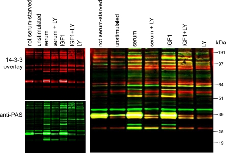
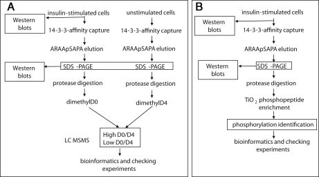
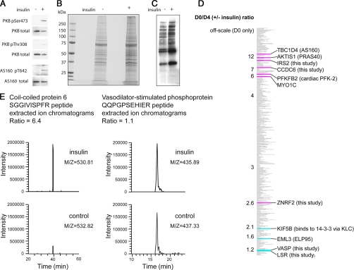
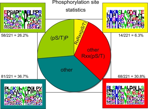
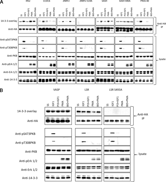
We devised a strategy of 14-3-3 affinity capture and release, isotope differential (d(0)/d(4)) dimethyl labeling of tryptic digests, and phosphopeptide characterization to identify novel targets of insulin/IGF1/phosphatidylinositol 3-kinase signaling. Notably four known insulin-regulated proteins (PFK-2, PRAS40, AS160, and MYO1C) had high d(0)/d(4) values meaning that they were more highly represented among 14-3-3-binding proteins from insulin-stimulated than unstimulated cells. Among novel candidates, insulin receptor substrate 2, the proapoptotic CCDC6, E3 ubiquitin ligase ZNRF2, and signaling adapter SASH1 were confirmed to bind to 14-3-3s in response to IGF1/phosphatidylinositol 3-kinase signaling. Insulin receptor substrate 2, ZNRF2, and SASH1 were also regulated by phorbol ester via p90RSK, whereas CCDC6 and PRAS40 were not. In contrast, the actin-associated protein vasodilator-stimulated phosphoprotein and lipolysis-stimulated lipoprotein receptor, which had low d(0)/d(4) scores, bound 14-3-3s irrespective of IGF1 and phorbol ester. Phosphorylated Ser(19) of ZNRF2 (RTRAYpS(19)GS), phospho-Ser(90) of SASH1 (RKRRVpS(90)QD), and phospho- Ser(493) of lipolysis-stimulated lipoprotein receptor (RPRARpS(493)LD) provide one of the 14-3-3-binding sites on each of these proteins. Differential 14-3-3 capture provides a powerful approach to defining downstream regulatory mechanisms for specific signaling pathways.


Figures

Fig. 1.
Fig. 1.
Proteins that bind to 14-3-3s in response to IGF1 in a PI 3-kinase-dependent manner. HEK293 cells cultured on 10-cm-diameter dishes in medium containing 10% (v/v) serum (labeled not serum-starved) were serum-starved for 4 h (unstimulated) and then stimulated as indicated with IGF1 at 50 ng/ml for 15 min and serum at 10% (v/v) for 15 min. Where indicated, cells were incubated with LY294002 (LY; 100 mm for 1 h) prior to stimulation. Cells were lysed in 0.3 ml/dish ice-cold lysis buffer, and 3 mg of each extract was added to 100 μl of a 50% (v/v) slurry of 14-3-3-Sepharose and mixed end over end for 4 h. After washing, the protein bound to 14-3-3-Sepharose was extracted into SDS sample buffer, separated by SDS-PAGE using a 4–12% gradient gel, and analyzed by Far-Western 14-3-3 overlay (red), which detects proteins on the blot that can bind directly to digoxigenin-labeled 14-3-3 proteins. Western blotting was also performed with the anti-PAS antibody (green).
Fig. 2.
Fig. 2.
Experimental strategy for identifying proteins whose phosphorylation and binding to 14-3-3s is stimulated by insulin. A, as detailed under “Experimental Procedures,” HeLa suspension cell cultures were serum-starved and stimulated or not with insulin. Proteins were captured on a 14-3-3-Sepharose column, which was washed until protein was undetectable in the flow-through, and specifically bound proteins were eluted by competition with a 1 mm concentration of the 14-3-3-binding synthetic phosphopeptide ARAApSAPA. Eluates were concentrated, digested with trypsin, and incubated with formaldehyde containing no deuterium (d0 for the preparation from insulin-stimulated cells) or two deuteriums (to give d4 for each dimethyl group added onto the peptides from unstimulated cells). d0/d4 ratios were determined as outlined under “Experimental Procedures.” B, strategy for identifying phosphopeptides derived from proteins isolated by 14-3-3 capture and release from extracts of insulin-stimulated cells. Phosphopeptides were enriched by titanium dioxide affinity as detailed further under “Experimental Procedures.”
Fig. 3.
Fig. 3.
d0/d4 (± insulin) ratios of 14-3-3 affinity-purified proteins from unstimulated and insulin-stimulated cells. A, comparison of PKB/Akt and AS160 total protein levels and phosphorylation status in lysates of unstimulated and insulin-stimulated HeLa cells used for 14-3-3 phosphoproteomics screens. B, Coomassie-stained SDS gel of proteins isolated by 14-3-3 capture and release from extracts of unstimulated and insulin-stimulated cells (5 times these amounts were run on a parallel gel and used for the MS analyses). C, 14-3-3 Far-Western overlay assay of proteins isolated by 14-3-3 capture and release from extracts of unstimulated and insulin-stimulated cells. D, thumbnail of the d0/d4 (±insulin) rankings for 14-3-3 affinity-purified proteins where off-scale indicates the proteins that were only detected in the preparation from insulin-stimulated cells. Highlighted in pink are proteins already known to be phosphorylated and to bind to 14-3-3s in response to insulin/IGF1 as well as proteins that were discovered to be insulin/IGF1-responsive in this study. In blue are proteins that we already knew do not respond to insulin/IGF1 as well as proteins found not to be insulin/IGF1-responsive in this study (see text). E, examples of d0/d4 ratio quantification for peptides from CCDC6 (d0/d4 = 6.4 for this peptide, contributing to the overall ratio of 7.1 for three quantified peptides from this protein (Table I)) and VASP (d0/d4 = 1.1 for this peptide with an overall ratio of 1.3 for the protein (supplemental Table 1)) showing extracted ion chromatograms after analysis in the LTQ-Orbitrap mass spectrometer.
Fig. 4.
Fig. 4.
Phosphopeptides derived from 14-3-3-binding proteins isolated from insulin-stimulated cells. The phosphorylated sites were classified according to whether they are found within motifs that conform to RXRXX(pS/pT), RXX(pS/pT) other than RXRXX(pS/pT), (pS/pT)P, or none of these (other). One phosphorylated site that matches both RXX(pS/pT) and (pS/pT)P is included in the RXX(pS/pT) group.
Fig. 5.
Fig. 5.
Cellular regulation of 14-3-3 binding of IRS2, CCDC6, ZNRF2, SASH1, PRAS40, VASP, and LSR. HA-IRS2, HA-CCDC6, HA-ZNRF2 (wild type and S19A mutant), HA-SASH1 (wild type and S90A mutant), HA-PRAS40, HA-VASP, and HA-LSR (wild type and S493A mutant) were isolated from transfected HEK293 cells that were stimulated as indicated, and anti-HA immunoprecipitates (IP) were analyzed by 14-3-3 overlay and Western blotting. Note that at higher exposures and in other experiments it was clear that 14-3-3 binding is not completely abolished with the ZNRF2 S19A mutation. As controls for the efficacy of stimuli and inhibitors, lysates were analyzed with antibodies against phospho-Thr308 and phospho-Ser473 of PKB/Akt, total PKB/Akt, phospho-Erk1/2 (pErk), and total Erk1/2. PI3, PI-103; BID, BI-D1870.

References

    1. Cohen P. (2006) The twentieth century struggle to decipher insulin signalling. Nat. Rev. Mol. Cell Biol. 7, 867–873 - PubMed
    1. Randhawa R., Cohen P. (2005) The role of the insulin-like growth factor system in prenatal growth. Mol. Genet. Metab. 86, 84– 90 - PubMed
    1. Manning B. D., Cantley L. C. (2007) AKT/PKB signaling: navigating downstream. Cell 129, 1261– 1274 - PMC - PubMed
    1. Hawkins P. T., Anderson K. E., Davidson K., Stephens L. R. (2006) Signalling through Class I PI3Ks in mammalian cells. Biochem. Soc. Trans. 34, 647– 662 - PubMed
    1. Yuan T. L., Cantley L. C. (2008) PI3K pathway alterations in cancer: variations on a theme. Oncogene 27, 5497–5510 - PMC - PubMed

Publication types