. 2009 Sep;10(9):1008-17.
doi: 10.1038/ni.1753. Epub 2009 Aug 2.

HIV-1 evades virus-specific IgG2 and IgA responses by targeting systemic and intestinal B cells via long-range intercellular conduits

Affiliations

HIV-1 evades virus-specific IgG2 and IgA responses by targeting systemic and intestinal B cells via long-range intercellular conduits

Weifeng Xu et al. Nat Immunol. 2009 Sep.

Abstract

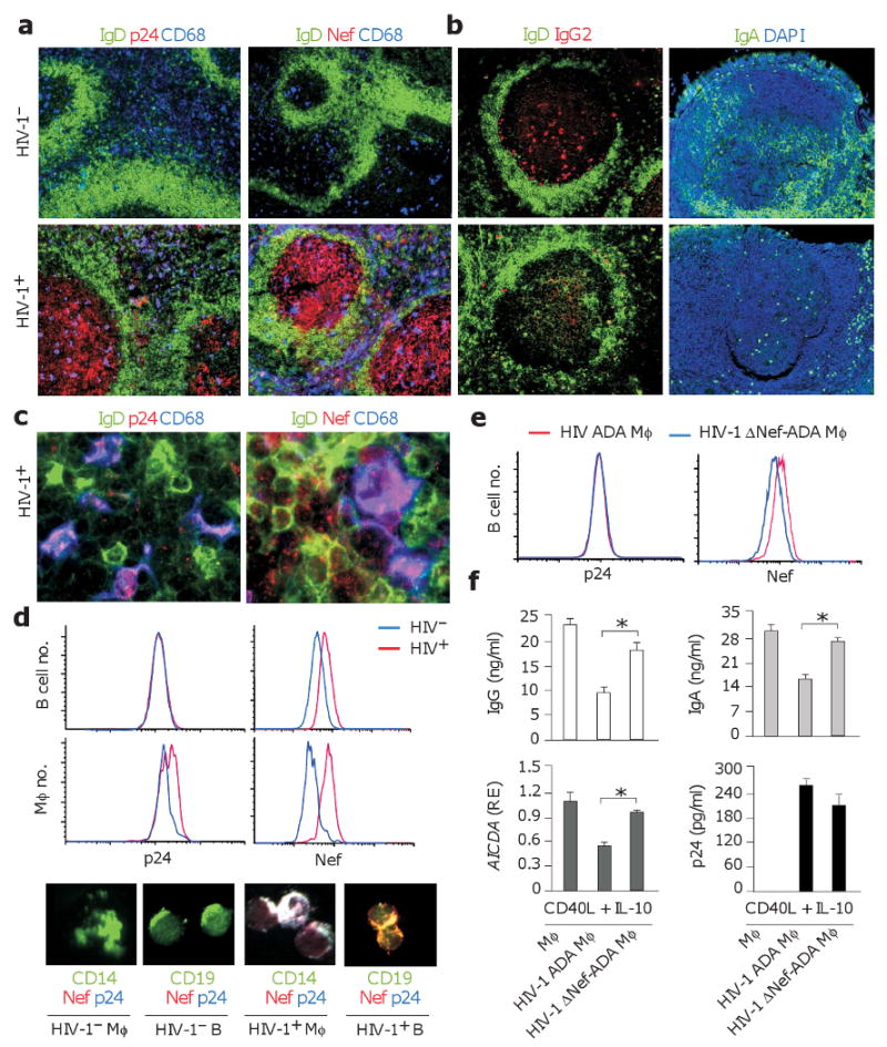
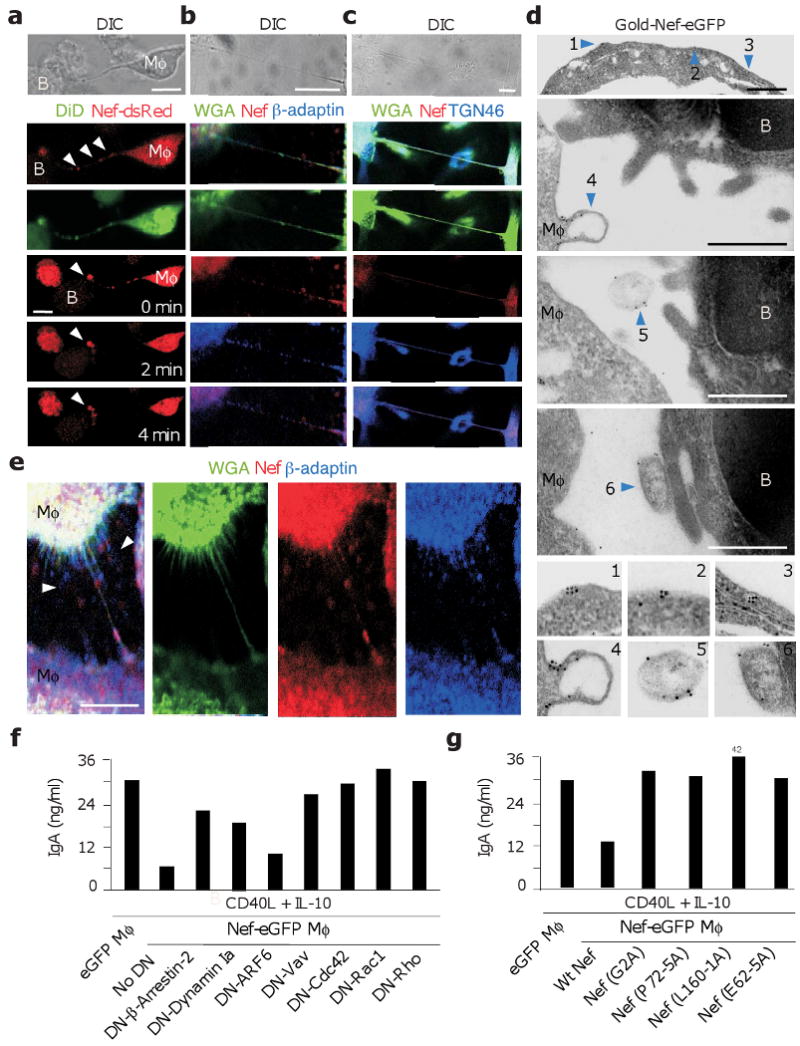
Contact-dependent communication between immune cells generates protection but also facilitates viral spread. Here we found that macrophages formed long-range actin-propelled conduits in response to negative factor (Nef), a human immunodeficiency virus type 1 (HIV-1) protein with immunosuppressive functions. Conduits attenuated immunoglobulin G2 (IgG2) and IgA class switching in systemic and intestinal lymphoid follicles by shuttling Nef from infected macrophages to B cells through a guanine-exchange factor-dependent pathway involving the amino-terminal anchor, central core and carboxy-terminal flexible loop of Nef. By showing stronger virus-specific IgG2 and IgA responses in patients with Nef-deficient virions, our data suggest that HIV-1 exploits intercellular 'highways' as a 'Trojan horse' to deliver Nef to B cells and evade humoral immunity systemically and at mucosal sites of entry.


Conflict of interest statement

Competing Interest Statement: The authors declare that they have no competing financial interests.

Figures

Figure 1
Figure 1. Infected primary macrophages transfer Nef to B cells and inhibit TD class switching
(a, b) Immunohistology of HIV-1 and HIV-1+ follicles from systemic lymph nodes or intestinal Peyer's patches stained for IgD or IgA (green), p24, Nef or IgG2 (red), and CD68 (blue). Original magnification, ×10. (c) Primary macrophages and B cells from HIV-1+ lymph nodes stained for IgD (green), p24 or Nef (red), and CD68 (blue). Original magnification, ×63. (d) Flow cytometry of p24 and Nef in primary CD19+ B cells and CD14+ macrophages (Mφ) sorted from HIV-1 (blue histograms) or HIV-1+ (red histograms) spleens. Similar cells were analyzed by immunofluorescence analysis upon staining for CD19 or CD14 (green), Nef (red), and p24 (blue). Original magnification, ×40. (e) p24 and Nef in CD19+ B cells sorted from a 24-h co-culture with primary macrophages infected with either wt ADA HIV-1 (red histograms) or ΔNef-ADA HIV-1 (blue histograms). (f) QRT-PCR of AICDA transcripts and ELISAs of IgG and IgA from IgD+ B cells co-cultured with primary uninfected, wt ADA-infected or ΔNef-ADA-infected macrophages in the presence of CD40L and IL-10 for 7 d. AICDA mRNA was quantified in B cells sorted after 4 d and normalized to ACTB mRNA. Relative expression (RE) compared to B cells stimulated with CD40L and IL-10 in the absence of macrophages. ELISA of p24 was performed 7 d after mock or ADA infection. Panels a-e show 1 of 5 experiments yielding similar results, whereas panel f summarizes 3 experiments (bars indicate mean s.d.; asterisk is p < 0.05).
Figure 2
Figure 2. Nef is sufficient for macrophage-like cells to acquire class switch-inhibiting functions
(a) Imaging of CellTracker-loaded IgD+ B cells (blue) co-cultured with eGFP-THP-1 or Nef-eGFP-THP-1 macrophage-like cells (Mφ, green) for 24 h. Original magnification, ×5. (b) Time course analysis of eGFP-THP-1 or Nef-eGFP-THP-1 in IgD+ B cells cultured as in a for 48 h. (c-e) QRT-PCR of Iα1-Cα1, Iα2-Cα2, AICDA and Iα-Cμ transcripts from IgD+ B cells cultured as in b, sorted and then incubated with or without CD40L and IL-10 for additional 48 h. Iα1-Cα1, Iα2-Cα2 and AICDA mRNAs were normalized to ACTB mRNA and Iα-Cμ mRNA to Iμ-Cμ mRNA. RE, relative expression. (f) ELISAs of IgG, IgA and IgM proteins from IgD+ B cells cultured as in c-e, except that the activation step was carried out for 7 days. (g) IgG, IgA and IgM proteins from IgD+ B cells incubated with supernatants from eGFP-THP-1- or Nef-eGFP-THP-1 cells in the presence or absence of CD40L and IL-10 for 7 days. (h, i) Immunohistology of HIV-1 and HIV-1+ follicles from systemic lymph nodes and intestinal Peyer's patches stained for IgD (green), AID (red), and Nef (blue or dark gray). DAPI (blue) counterstains nuclei. Original magnification, ×10. Panels a, h and i show 1 of 5 experiments yielding similar results, whereas panels b-g summarize 3 experiments (bars indicate mean s.d.; asterisk is p < 0.05). Scale bar equals 10 μm.
Figure 3
Figure 3. Nef utilizes multiple motifs to stimulate conduit formation in macrophage-like cells
(a) Live-cell confocal imaging of THP-1 macrophage-like cells (Mφ) 24 h after nucleofection with eGFP (control), wt Nef-eGFP, Nef (G2A)-eGFP, Nef (P72-5A)-eGFP, Nef (L160-1A)-eGFP or Nef (E62-5A)-eGFP plasmid. (b) Live-cell confocal imaging of Nef-eGFP-THP-1 cells (green) co-cultured with CellTracker-loaded IgD+ B cells (red) for 6 h. Bottom panels show Nef-eGFP-THP-1 cells after three-dimensional reconstruction. (c) Average number of protrusions on individual THP-1 cells nucleofected as in A (upper panel), and percentage of IgD+ B cells acquiring eGFP fluorescence after co-culture with THP-1 cells nucleofected as in A for 24 h. (d) Live-cell DIC (left and mid panels) and confocal (right panel) imaging of Nef-eGFP-THP-1 cells (green) co-cultured with CellTracker-loaded IgD+ B cells (red). Dashed box in left panel includes cells magnified in mid panel. (e) Live-cell DIC imaging of THP-1 cells pre-loaded with LysoTracker and co-cultured with IgD+ B cells. In upper panel, arrowheads show a long-range tubular conduit containing CellTracker, whereas dashed rectangle pinpoints short-range transfer of CellTracker from a THP-1 cell to a B cell. In bottom panels, short-range transfer is shown over time, each panel being separated by approximately 15 sec. Panels a, b, d and e show 1 of 4 experiments yielding similar results, whereas panel c summarizes 3 experiments (bars indicate mean s.d.; asterisk is p < 0.05 compared to wt Nef-eGFP value). Scale bar equals 10 μm.
Figure 4
Figure 4. Infected primary macrophages transfer Nef to B cells via long-range actin-propelled conduits
(a) Confocal imaging of uninfected, wt ADA-infected or ΔNef-ADA-infected macrophages (Mφ) stained for Nef (red) or p24 (blue) in the presence of WGA (green). Leftmost panel shows number of long (> 100 μm) tethers/50 uninfected or infected macrophages. Original magnification, ×40. (b-d) ADA-infected macrophages cultured with or without IgD+ B cells and stained for Nef (red) and p24 or Pax5 (blue) in the presence of WGA (green). Original magnification, ×5 (b and c, upper panels), ×63 (b and c, lower panels), ×10 (d). (e, f) Percentage of THP-1-Nef-eGFP macrophage-like cells (Mφ) forming tethers and percentage of IgD+ B cells acquiring Nef-eGFP from Nef-eGFP-THP-1 cells in the presence or absence of chemical inhibitors (azide, latrunculin B, colchicin, nocodazole, wortmannin, AD101 or ADMD3100) or DN inhibitors (to β-arrestin-2, dynamin Ia, Arf6, Vav2, Cdc42, Rac1 or Rho). Control value corresponds to no inhibitor or empty construct and was set at 100%. Panels a-d show 1 of 4 experiments yielding similar results, whereas panels e and f summarize 3 experiments (bars indicate mean s.d.; asterisk is p < 0.05 compared to control). Scale bar equals 10 μm.
Figure 5
Figure 5. Macrophage-like cells inhibit class switching by trafficking membrane and vesicular Nef via Vav and small GTPases
(a) Live DIC (uppermost panels) or confocal imaging of Nef-dsRed-THP-1 macrophage-like cells (Mφ, red) co-stained with the membrane-specific probe DiD (green) and incubated with unstained IgD+ B cells. Arrowheads point to organelle-like beads within an intercellular conduit. The bottom three panels from the top show intercellular transfer of Nef-dsRed over time. Arrowheads point to site of Nef-dsRed accumulation in a B cell. Original magnification, ×40. (b, c) Confocal imaging of ADA-infected primary macrophages (Mφ) stained for Nef (red) and β-adaptin or TGN46 (blue) in the presence of WGA (green). (d) Transmission electron microscopy of Nef-eGFP-THP-1 macrophage-like cells (Mφ) co-cultured with IgD+ B cells for 6 hours and stained with a gold-labeled monoclonal antibody to GFP. Arrowheads 1 and 2, membrane-bound Nef on a Mφ-derived conduit; arrowhead 3, Nef within vesicular and tubular structures from a Mφ-derived conduit; arrowheads 4-6, Nef associated with Mφ budding as well as free or B cell-docked exosome-like bodies, respectively. Original magnification, ×36000. (e) Confocal imaging of ADA-infected primary macrophages stained for Nef (red) or β-adaptin (blue) in the presence of WGA (green). Arrowheads point to extracellular bodies. (f) ELISAs of IgA from IgD+ B cells incubated with THP-1-Nef-eGFP cells nucleofected with either an empty plasmid (control) or a DN plasmid to β-arrestin-2, dynamin Ia, ARF6, Vav, Cdc42, Rac1 or Rho. After 48 h, B cells were sorted and cultured for additional 7 days with or without CD40L and IL-10. (g) ELISAs of IgA from IgD+ B cells incubated with THP-1 cells expressing eGFP, wt Nef-eGFP, Nef (G2A)-eGFP, Nef (P72-5A)-eGFP, Nef (L160-1A)-eGFP or Nef (E62-5A)-eGFP. B cells were cultured as in f. Panels a-g show 1 of 4 experiments yielding similar results. Scale bars equal 10 and 1 μm in confocal and transmission electron microscopy images, respectively.
Figure 6
Figure 6. Nef inhibits class switching in GCs but not extrafollicular areas
(a, b) Immunohistology of HIV-1 and HIV-1+ follicles from systemic lymph nodes stained for IgD (green), Nef (red), and CD11c (blue). DAPI (blue) counterstains nuclei. Arrowheads point to intercellular conduits. Mφ, macrophages. Original magnification, ×63. (c-e) HIV-1 and HIV-1+ follicles from systemic lymph nodes or intestinal Peyer's patches stained for IgD or IgA (green), AID, ferritin or BAFF (red), and Nef or CD68 (blue). DAPI (blue) counterstains nuclei. Original magnification, ×10. (f) ELISA of ferritin from uninfected or ADA-infected primary macrophages. (g) ELISAs of IgM, IgG and IgA from IgD+ B cells co-cultured with CD40L or BAFF and IL-10 in the presence or absence of ferritin for 7 d. Panels a-e show 1 of 4 experiments yielding similar results, whereas panels f and g summarize 3 experiments. Scale bar equals 10 μm.
Figure 7
Figure 7. ΔNef HIV-1 elicits more virus-specific IgG2, IgA1 and IgA2 than wt HIV-1 irrespective of viral load
(a) Serum IgG2, IgG3, IgA1 and IgA2 to viral p24 Gag, Tat and gp120 Env in ΔNef HIV-1+ LTNPs and wt HIV-1+ LTNPs. Each value from ΔNef HIV-1+ LTNPs represents the average of 3 to 5 independent determinations performed over a time period ranging from 6 to 12 years as indicated in Supplementary Table 2 online. Values are expressed as optical density (OD) at 450 nm. (b) Linear regression analysis of viral load and p24-specific IgA1 in D36, C54, C64 and C98 ΔNef HIV-1+ LTNPs examined at multiple time points as indicated in Supplementary Table S2.

References

    1. Davis DM. Intercellular transfer of cell-surface proteins is common and can affect many stages of an immune response. Nat Rev Immunol. 2007;7:238–243. - PubMed
    1. Rustom A, Saffrich R, Markovic I, Walther P, Gerdes HH. Nanotubular highways for intercellular organelle transport. Science. 2004;303:1007–1010. - PubMed
    1. Sherer NM, Mothes W. Cytonemes and tunneling nanotubules in cell-cell communication and viral pathogenesis. Trends Cell Biol. 2008;18:414–420. - PMC - PubMed
    1. Watkins SC, Salter RD. Functional connectivity between immune cells mediated by tunneling nanotubules. Immunity. 2005;23:309–318. - PubMed
    1. Fackler OT, Alcover A, Schwartz O. Modulation of the immunological synapse: a key to HIV-1 pathogenesis? Nat Rev Immunol. 2007;7:310–317. - PubMed

Publication types

MeSH terms