Clinical Trial
. 2009 Aug;15(8):886-92.
doi: 10.1038/nm.2006. Epub 2009 Aug 2.

Persistence of HIV-1 receptor-positive cells after HSV-2 reactivation is a potential mechanism for increased HIV-1 acquisition

Affiliations
Clinical Trial

Persistence of HIV-1 receptor-positive cells after HSV-2 reactivation is a potential mechanism for increased HIV-1 acquisition

Jia Zhu et al. Nat Med. 2009 Aug.

Abstract

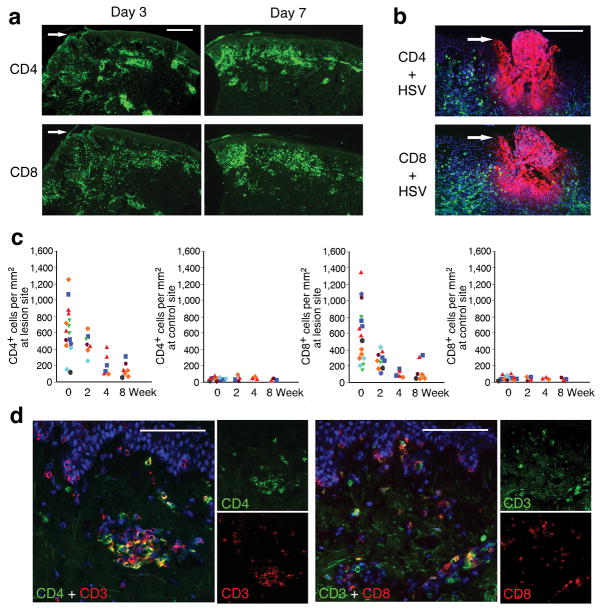
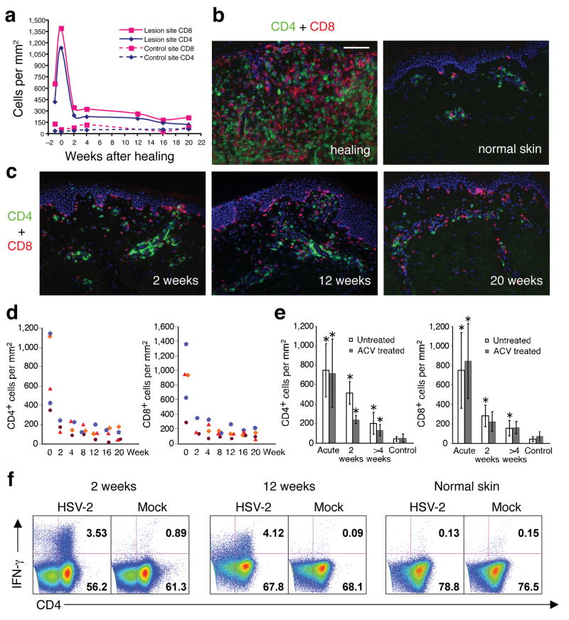
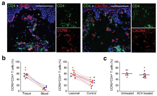
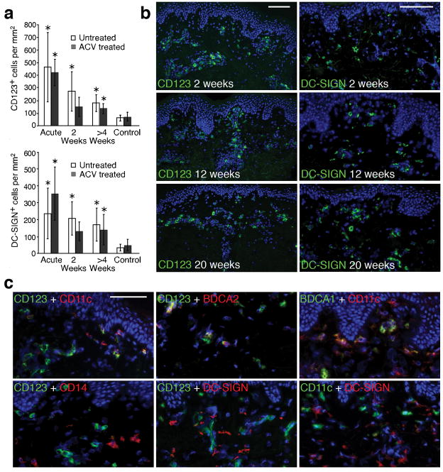
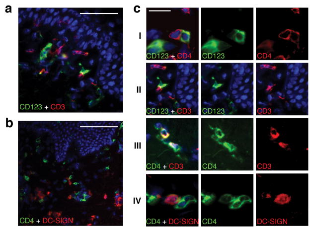
To explore the mechanism by which herpes simplex virus (HSV)-2 infection is related to HIV-1 acquisition, we conducted in situ analysis of the cellular infiltrate from sequential biopsies of HSV-2 lesions from patients on and off antiviral therapy. CD4(+) and CD8(+) T cells and a mixed population of plasmacytoid and myeloid dendritic cells (DCs), including cells expressing the C-type lectin receptor DC-SIGN, persisted at sites of HSV-2 reactivation for months after healing, even with daily antiviral therapy. The CD4(+) T cells that persisted reacted to HSV-2 antigen, were enriched for expression of the chemokine receptor CCR5, and were contiguous to DCs expressing the interleukin-3 receptor CD123 or DC-SIGN. Ex vivo infection with a CCR5-tropic strain of HIV-1 revealed greater concentrations of integrated HIV-1 DNA in cells derived from healed genital lesion biopsies than in cells from control skin biopsies. The persistence and enrichment of HIV receptor-positive inflammatory cells in the genitalia help explain the inability of anti-HSV-2 therapy to reduce HIV acquisition.


Figures

Figure 1
Figure 1. Persistence and localization of CD4+ and CD8+ T cells in genital lesions of immunocompetent subjects
(a) Overview of infiltrating CD4+ and CD8+ cells in skin biopsies of an acute ulcer (d 3) and recently healed lesion (d 7) taken from a single subject who reactivated HSV-2 at two distinct anatomic sites. The arrows illustrate the area of epithelial ulceration. (b) Infiltration of CD4+ (top) and CD8+ (bottom) cells near the site of HSV-2 (red) replication (d 2 lesion) in another patient. (c) Quantitation of CD4+ and CD8+ cells in skin biopsies from the lesion site as compared to uninvolved (control) genital skin biopsies. At least five fields from each biopsy were counted for defining the density of cells . Each symbol represents one subject (n = 8). Biopsies from acute herpetic lesion, including the ulcerative (d 1–4) and newly healed (d 6–8) stages, were collated into the week 0 time point. (d) Dual immunofluorescence staining with CD3 and CD4 or CD8 of biopsy after complete healing of the lesion site. Scale bar: 400 μm (a), 200 μm (b), and 100 μm (d).
Figure 2
Figure 2. Persistence of CD4+ and CD8+ T cells in genital skin of subjects on daily acyclovir therapy
(a) Quantitation of CD4+ and CD8+ cells infiltrating an HSV-2 lesion vs. uninvolved (control) genital skin. Week 0 is the time of complete lesion healing (lesion onset is labeled week -1). (b and c) Dual immunofluorescence staining of CD4+ (green) and CD8+ (red) cells. Panel b: comparison of healing stage at lesion site versus normal uninvolved genital skin. Panel c: shows CD4+ and CD8+ cells in post-healing biopsies taken from the time points indicated in panel a. (d) Quantitation of CD4+ and CD8+ cells in genital lesions of all four subjects on acyclovir therapy from acute lesion to 20 weeks post-healing. Each symbol represents a subject. (e) Comparison of CD4+ and CD8+ T cell infiltration in genital skin from untreated versus acyclovir-treated HSV recurrence. No statistically significant differences were present between acyclovir versus untreated episodes either during or after herpes reactivation (P > 0.5). The asterisk marks biopsies of statistical significance (P < 0.05) in comparison of lesion site versus control site. (f) Persistent CD4+ T cells in post-healing biopsies were HSV-2 antigen specific. After incubation with whole UV-killed HSV-2 antigen, IFN-γ expression was detected in lymphocytes collected from biopsies at 2 and 12 weeks post-HSV-2 reactivation but not in lymphocytes collected from the uninvolved genital skin (12 weeks post healing) of the subject depicted in panels a-c. Scale bar: 100 μm (B and C).
Figure 3
Figure 3. HIV co-receptor expression on CD4+ T cells in HSV-2 lesions
(a) CCR5 and CXCR4 expression on CD4+ cells in lesions after healing. A 2 weeks post healing biopsy from an acyclovir treated subject was dual stained with either CCR5 and CD4, or CXCR4 and CD4 antibodies. (b) Comparison of CCR5 expression on CD4+ cells in genital skin after infection (n = 20) and in circulating PBMC (n = 11) (left panel) or in unaffected control skin (n = 15) (right panel). Each symbol represents one subject. Lines connect samples taken from the same subject on the same day (p=0.04 for comparison in number of CCR5-expressing CD4+ cells from genital skin previously infected with HSV-2 versus contralateral non-infected genital skin). (c) Comparison of CCR5 expression in post-healing biopsies during acyclovir-treated (n = 12) versus untreated (n = 8) time periods (P > 0.5). Lines indicate the median percentage of CCR5-expressing CD4+ cells. Scale bar: 100 μm.
Figure 4
Figure 4. Persistence of dendritic cells in genital lesion biopsies
(a) Quantitation of CD123+ and DC-SIGN+ cells in lesion-site biopsies during acyclovir-treated and untreated time periods. An asterisk above a bar denotes that the number of CD123+ or DC-SIGN+ cells in a lesion-site skin biopsy was significantly higher than in control skin (P < 0.05). Acyclovir had no statistically significant effect on the persistence of CD123+ or DC-SIGN+ DCs over time (p=0.96 and p=0.64, respectively). (b) Representative skin biopsy sections demonstrating the persistence of DCs positive for either CD123 or DC-SIGN in the upper dermis and near blood vessels in post-healing biopsies obtained at 2, 12 and 20 weeks while on daily acyclovir. (c) Detection of a diverse DC population in post-healing biopsies by co-staining of CD123 or DC-SIGN with CD11c, CD14, BDCA1 and BDCA2 in post-healing biopsies (week 16–20) during acyclovir treatment. The top first panel shows that three populations of DCs: CD123+CD11c, CD123+CD11c+, and CD123CD11c+. The top second panel shows co-staining of CD123 and BDCA2 and the top third panel shows co-staining of the CD11c+ cells with BDCA1. The bottom first panel shows the lack of CD14 expression on CD123+ cells. The bottom second and third panel demonstrate that DC-SIGN expressing cells do not co-express CD123 or CD11c. Scale bar: 100 μm (b), 50 μm (c).
Figure 5
Figure 5. Interactions of DCs and CD4+ T cells in post-healing biopsies during acyclovir treatment
(a) Dual immunofluorescence staining of CD123 with CD3 showing the interaction of CD123+ cells (green) and CD3+ T cells (red) in a week 12 post-healing biopsy while on acyclovir therapy. (b) Associations of CD4+ cells (green) with cells expressing DC-SIGN (red). Biopsy was taken after 20 weeks of acyclovir therapy. (c) Detailed micrographs illustrate direct interaction between CD123+ DCs and CD4+ T cells, and DC-SIGN+ DCs and CD4+ T cells at genital lesion site post healing. Panel I shows CD123+ DC are contiguous to CD4+ T cells. Panel II shows interactions between CD123+ DC cells and T cells. Panel III illustrates clusters of CD3+CD4high T cells and CD3-CD4low DCs. Panel IV shows a CD4+ T cell contiguous to a DC-SIGN expressing cell scale bar:50μ(a), 100μ(b), 20μ(c).

References

    1. Freeman EE, et al. Herpes simplex virus 2 infection increases HIV acquisition in men and women: systematic review and meta-analysis of longitudinal studies. Aids. 2006;20:73–83. - PubMed
    1. Wald A, Link K. Risk of Human Immunodeficiency Virus (HIV) Infection in Herpes Simplex Virus Type-2 (HSV-2) Seropositive Persons: a Meta-Analysis. J Infect Dis. 2002;185:45–52. - PubMed
    1. Koelle D, et al. Antigenic specificities of human CD4+ T-cell clones recovered from recurrent genital herpes simplex virus type 2 lesions. J Virolo. 1994;68:2803–2810. - PMC - PubMed
    1. Wald A, et al. Frequent genital HSV-2 shedding in immunocompetent women. J Clin Invest. 1997;99:1092–1097. - PMC - PubMed
    1. Gupta R, et al. Valacyclovir and acyclovir for suppression of shedding of herpes simplex virus in the genital tract. J Infect Dis. 2004;190:1374–1381. - PubMed

Publication types

MeSH terms