

Review
doi: 10.1038/nrmicro2171.
Epub 2009 Aug 3.
Listeria monocytogenes - from saprophyte to intracellular pathogen
Affiliations
- PMID: 19648949
- PMCID: PMC2813567
- DOI: 10.1038/nrmicro2171
Item in Clipboard
Review
Listeria monocytogenes - from saprophyte to intracellular pathogen
Nat Rev Microbiol.
2009 Sep.
Abstract
Listeria monocytogenes is a bacterium that lives in the soil as a saprophyte but is capable of making the transition into a pathogen following its ingestion by susceptible humans or animals. Recent studies suggest that L. monocytogenes mediates its saprophyte-to-cytosolic-parasite transition through the careful modulation of the activity of a virulence regulatory protein known as PrfA, using a range of environmental cues that include available carbon sources. In this Progress article we describe the regulation of PrfA and its role in the L. monocytogenes transition from the saprophytic stage to the virulent intracellular stage.
Figures

Listeria monocytogenes survives in a diverse array of environments, in habitats that include soil and water as well as food processing facilities. Central to the switch between life outside and life inside mammalian hosts is the transcriptional activator PrfA, which regulates the expression of many gene products that are required for bacterial virulence. Outside a host cell, PrfA exists in a low-activity state, with correspondingly low levels of virulence gene expression. Once inside the host, PrfA becomes activated (PrfA*) and induces the expression of gene products that are needed for host cell invasion (internalins InlA and InlB), phagosome lysis (listeriolysin O (LLO), phosphatidylinositol-specific phospholipase C (PI-PLC) and phosphatidylcholine (PC)-PLC), intracellular growth (hexose-6-phosphate transporter (Hpt)), and cell-to-cell spread (actin assembly-inducing protein (ActA); actin polymerization is shown in turquoise). The intracellular life cycle is modified, with permission, from REF. 81© (1989) Rockefeller University Press.

The phosphoenol pyruvate (PEP) transport system (PTS) is a multiprotein phosphorelay system that couples the transport of sugars across the bacterial cell membrane with their simultaneous phosphorylation. The PTS is composed of three distinct proteins: enzyme I (EI), histidine protein (HPr) and enzyme II (EII). A separate and distinct transporter, Hpt, mediates the transport of hexose phosphates, such as glucose-6-phosphate. a | In the presence of PTS-dependent sugars, EI (which autophosphorylates using the phosphoryl group from PEP) transfers a phosphoryl group to HPr, which then transfers it to the A domains of the various substrate-specific transporters or EIIs. During sugar transport, the phosphoryl group of EIIA is rapidly transferred to the EIIB domain and, from there, to the incoming carbohydrate as it passes through the membrane. EIIA therefore exists primarily in a non-phosphorylated state during active PTS sugar transport, and it is this form of EIIA that is postulated to sequester PrfA and inhibit its activity. b | In the presence of non-PTS-dependent sugars, such as glucose-6-phosphate, transport occurs through an alternative transporter such as Hpt. The EIIA component of PTS remains phosphor-ylated and is unable to sequester PrfA in this state. PrfA is released and can directly activate target promoters. c | Alternatively, PrfA that is released from EIIA may subsequently require the additional stimulus of an activating signal or cofactor to fully induce the expression of PrfA-dependent promoters.
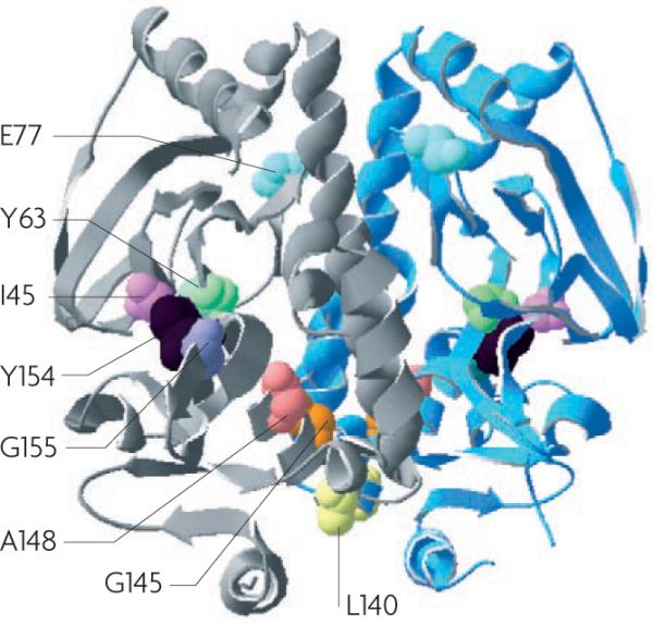
The crystal structure of a PrfA dimer (monomers are shown in grey and in light blue). The locations of the residues that are substituted in the PrfA* mutations described in the literature are as follows,,,: E77K, shown in light blue; Y63C, shown in light green; I45S, shown in pink; G155S, shown in purple; A148T, shown in salmon; G145S, G145R or G145C, shown in orange; L140F, shown in yellow. The Y154C mutation shown in black is unique in that it locks the protein into an activity state that enhances in vitro gene expression but does not provide full activation of PrfA-dependent gene expression in vivo. Figure is modified, with permission, from REF. 59 © (2008) Society for General Microbiology.
References
-
- Sakamoto M, Umeda M, Benno Y. Molecular analysis of human oral microbiota. J. Periodont. Res. 2005;40:277–285. - PubMed
-
- Zoetendal EG, Rajilic-Stojanovic M, de Vos WM. High-throughput diversity and functionality analysis of the gastrointestinal tract microbiota. Gut. 2008;57:1605–1615. - PubMed
-
- Gans J, Wolinsky M, Dunbar J. Computational improvements reveal great bacterial diversity and high metal toxicity in soil. Science. 2005;309:1387–1390. - PubMed
Publication types
MeSH terms
Substances
Grants and funding
LinkOut - more resources
Full Text Sources
Other Literature Sources
Medical
