. 2008 Oct;3(5):569-585.
doi: 10.1586/17469872.3.5.569.

Current concepts of metastasis in melanoma

Affiliations

Current concepts of metastasis in melanoma

Blazej Zbytek et al. Expert Rev Dermatol. 2008 Oct.

Abstract

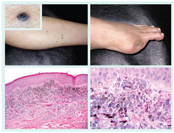
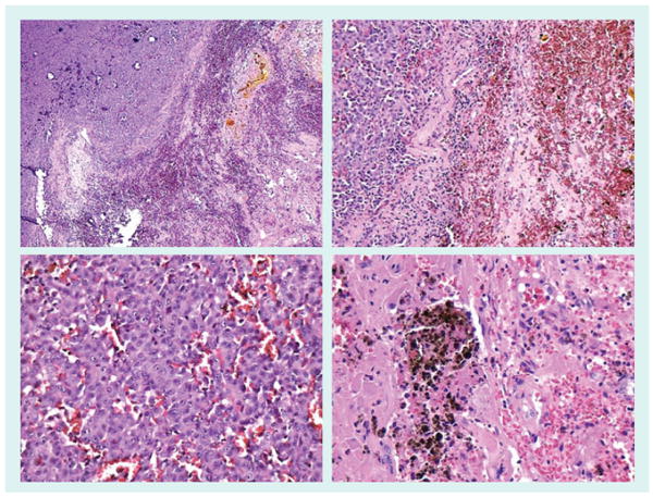
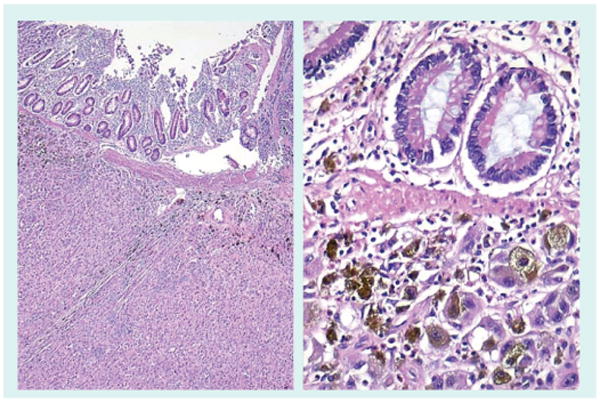
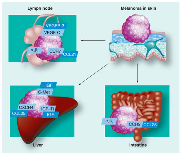
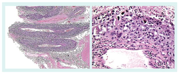
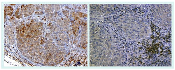
The main cause of death in melanoma patients is widespread metastases. Staging of melanoma is based on the primary tumor thickness, ulceration, lymph node and distant metastases. Metastases develop in regional lymph nodes, as satellite or in-transit lesions, or in distant organs. Lymph flow and chemotaxis is responsible for the homing of melanoma cells to different sites. Standard pathologic evaluation of sentinel lymph nodes fails to find occult melanoma in a significant proportion of cases. Detection of small numbers of malignant melanoma cells in these and other sites, such as adjacent to the primary site, bone marrow or the systemic circulation, may be enhanced by immunohistochemistry, reverse transcription PCR, evaluation of lymphatic vessel invasion and proteomics. In the organs to which melanoma cells metastasize, extravasation of melanoma cells is regulated by adhesion molecules, matrix metalloproteases, chemokines and growth factors. Melanoma cells may travel along external vessel lattices. After settling in the metastatic sites, melanoma cells develop mechanisms that protect them against the attack of the immune system. It is thought that one of the reasons why melanoma cells are especially resistant to killing is the fact that melanocytes (cells from which melanoma cells derive) are resistant to such noxious factors as ultraviolet light and reactive oxygen species. Targeted melanoma therapies are, so far, largely unsuccessful, and new ones, such as adjuvant inhibition of melanogenesis, are under development.


Figures

Figure 1
Figure 1. Breslows tumor thickness and ulceration are currently the American Joint Committee on Cancer criteria for staging of primary cutaneous melanoma and the most predictive factor for occult metastasis at the time of diagnosis
Figure 2
Figure 2. Histologic and clinical findings that classify the tumor as American Joint Committee on Cancer stage III
(A) Microscopic, immunohistochemistry positive (MART1) sentinel lymph node, (B) clinically and pathogically positive lymph node and (C) peritumoral and in transit metastasis (right).
Figure 3
Figure 3. Factors that are predictive of metastasis in the primary
(A) Mitotic rate, (B) vascular invasion, (C) absence of a tumor-infiltrating lymphocyte host response and (D) microsatellites, whose presence upgrades the melanoma to American Joint Committee on Cancer stage IIIc and are essentially in-transit metastasis.
Figure 4
Figure 4. Melanomas are characterized (similar to most metastatic tumors) as a heterogenous population of tumor cells that are genetically unstable and lead to clonal divergence and acquisition of metastatic phenotype
A desmoplastic melanoma that developed a dark papule that lead to clinical and pathologic recognition is shown. The patient developed a lung metastasis with a similar population of cells.
Figure 5
Figure 5. Melanoma metastatic to subcutaneous tissue
Distant subcutaneous melanoma metastases are a common phenomenon. Illustrated here is a well-circumscribed subcutaneous melanoma nodule from the scalp. Resection of distant metastases (metastatectomy), such as subcutaneous metastases, in selected melanoma patients is associated with improved 5-year survival [160].
Figure 6
Figure 6. In-transit melanoma metastasis of the lower extremity
Extremity melanomas are often associated with a protracted course characterized by in-transit metastasis slowly progressing to the regional lymph node basin and beyond. The metastases can be epidermotropic and mimic a primary melanoma. Amputation of an acral melanoma of the first toe (top right panel) was followed by the acquisition of numerous ascending in-transit metastases, seen as small dark papules (top left panel and inset). Biopsy revealed well-defined superficial dermal nodules (bottom left panel) of melanoma cells abutting the epidermis (bottom right panel).
Figure 7
Figure 7. Melanoma brain metastasis
Brain metastases are common in melanoma and often hemorrhagic [161]. The top left panel depicts a cerebral metastases (top left) bordering a necrotic and hemorrhagic area (bottom right). The bottom left panel shows extravasated red blood cells and small vessels surrounding a nest of melanoma cells. The top right panel shows cerebral metastases (left side) adjacent to a large area of hemorrhage (right side). The bottom right panel illustrates both hemosiderin and hemorrhage in brain parenchyma.
Figure 8
Figure 8. Melanoma lung metastasis
The lung is the most common sight of cutaneous melanoma visceral metastasis. Depicted in the left panel is replacement of most of the lung parenchyma by metastases. The top right panels shows the melanoma cells infiltrating alveolar spaces. The bottom right panel shows a metastasis with variable melanization of tumor cells.
Figure 9
Figure 9. Melanoma small intestine metastasis
Metastasis to small intestine are rare and most often due to melanoma. The left panel shows submucosal small intestinal metastases. The right panel illustrates a small cluster of melanoma cells in the lamina propria.
Figure 10
Figure 10. Selected mechanisms of chemotaxis of melanoma cells to lymph nodes and distant organs
Figure 11
Figure 11. Melanoma angiotropism
Angiotropism, perivascular cuffing by melanoma cells of the external surface of vessels, is a recently described phenomenon [162], which has been shown to independently predict for local recurrence and in-transit metastasis [163]. The left panel shows the expansion of the media and adventitia of several small muscular vessels by melanoma cells. The right panel demonstrates a small vessel surrounded by a cuff of melanoma cells.
Figure 12
Figure 12. Selection of therapy and phenotyping of melanoma metastases
Determination of the phenotype and genotype of metastatic melanoma may play a new role in pathology to aid in the selection of targeted therapy. The antiapoptotic protein Bcl-2 is suspected to influence the treatment responsiveness of melanoma. The lack of Bcl-2 expression demonstrated in the melanoma metastasis in the right panel (lymphocytes are internal positive controls) has been associated with a higher response rate to chemoimmunotherapy in comparison to a diffuse and focal pattern of Bcl-2 expression found in the melanoma metastasis depicted in the left panel [164].

Similar articles

Cited by

References

    1. Balch CM, Soong SJ, Atkins MB, et al. An evidence-based staging system for cutaneous melanoma. CA Cancer J Clin. 2004;54(3):131–149. - PubMed
    1. Balch CM, Buzaid AC, Soong SJ et al. Final version of the American Joint Committee on Cancer staging system for cutaneous melanoma. J Clin Oncol. 2001;19(16):3635–3648. - PubMed
    2. • Current melanoma staging system.

    1. Bastian BC, Kashani-Sabet M, Hamm H, et al. Gene amplifications characterize acral melanoma and permit the detection of occult tumor cells in the surrounding skin. Cancer Res. 2000;60(7):1968–1973. - PubMed
    1. North JP, Kageshita T, Pinkel D, Leboit PE, Bastian BC. Distribution and significance of occult intraepidermal tumor cells surrounding primary melanoma. J Invest Dermatol. 2008;128(8):2024–2030. - PMC - PubMed
    1. Xu X, Gimotty PA, Guerry D, et al. Lymphatic invasion revealed by multispectral imaging is common in primary melanomas and associates with prognosis. Hum Pathol. 2008;39(6):901–909. - PMC - PubMed

Website

    1. American Cancer Society; GA, USA: 2008. Cancer facts and figures 2007. www.cancer.org/downloads/STT/CAFF2007PWSecured.pdf.

LinkOut - more resources