. 2009 Jul 31;4(7):e6458.
doi: 10.1371/journal.pone.0006458.

Punicic acid a conjugated linolenic acid inhibits TNFalpha-induced neutrophil hyperactivation and protects from experimental colon inflammation in rats

Affiliations

Punicic acid a conjugated linolenic acid inhibits TNFalpha-induced neutrophil hyperactivation and protects from experimental colon inflammation in rats

Tarek Boussetta et al. PLoS One. .

Abstract

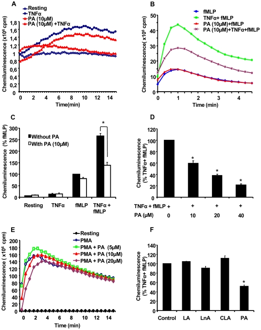
Background: Neutrophils play a major role in inflammation by releasing large amounts of ROS produced by NADPH-oxidase and myeloperoxidase (MPO). The proinflammatory cytokine TNFalpha primes ROS production through phosphorylation of the NADPH-oxidase subunit p47phox on Ser345. Conventional anti-inflammatory therapies remain partially successful and may have side effects. Therefore, regulation of neutrophil activation by natural dietary components represents an alternative therapeutic strategy in inflammatory diseases such as inflammatory bowel diseases. The aim of this study was to assess the effect of punicic acid, a conjugated linolenic fatty acid from pomegranate seed oil on TNFalpha-induced neutrophil hyperactivation in vitro and on colon inflammation in vivo.

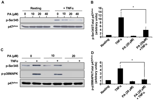
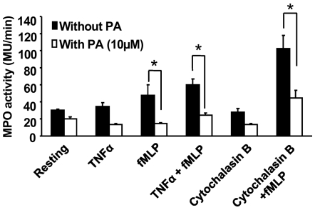
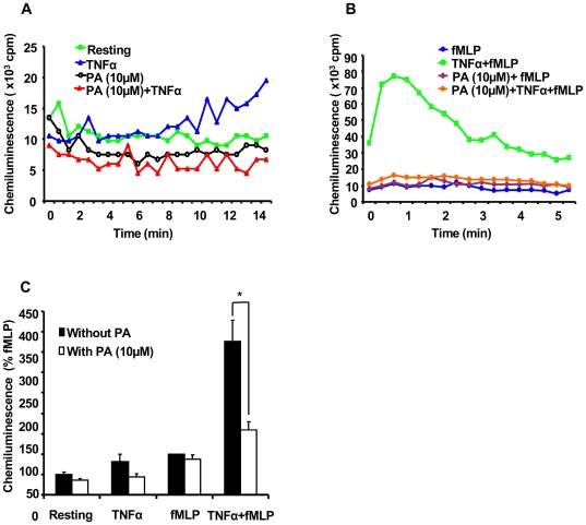
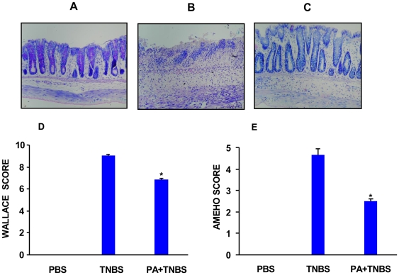
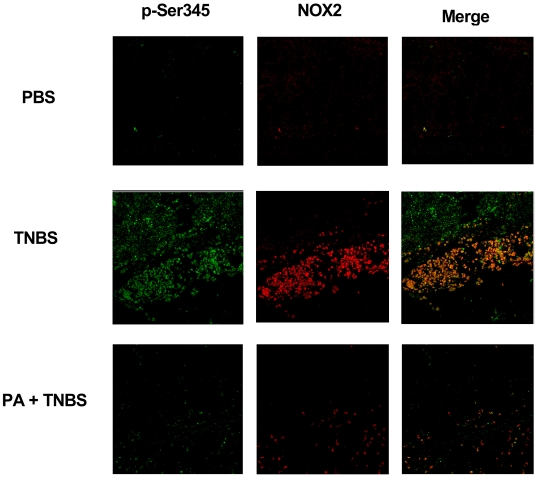
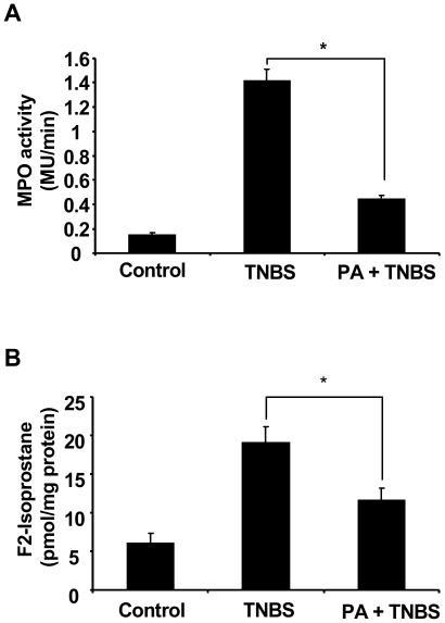
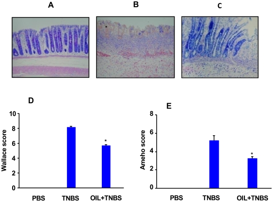
Methodology and principal findings: We analyzed the effect of punicic acid on TNFalpha-induced neutrophil upregulation of ROS production in vitro and on TNBS-induced rat colon inflammation. Results show that punicic acid inhibited TNFalpha-induced priming of ROS production in vitro while preserving formyl-methionyl-leucyl-phenylalanine (fMLP)-induced response. This effect was mediated by the inhibition of Ser345-p47phox phosphorylation and upstream kinase p38MAPK. Punicic acid also inhibited fMLP- and TNFalpha+fMLP-induced MPO extracellular release from neutrophils. In vivo experiments showed that punicic acid and pomegranate seed oil intake decreased neutrophil-activation and ROS/MPO-mediated tissue damage as measured by F2-isoprostane release and protected rats from TNBS-induced colon inflammation.

Conclusions/significance: These data show that punicic acid exerts a potent anti-inflammatory effect through inhibition of TNFalpha-induced priming of NADPH oxidase by targeting the p38MAPKinase/Ser345-p47phox-axis and MPO release. This natural dietary compound may provide a novel alternative therapeutic strategy in inflammatory diseases such as inflammatory bowel diseases.


Conflict of interest statement

Competing Interests: The authors have declared that no competing interests exist.

Figures

Figure 1
Figure 1. Punicic acid (PA) inhibits TNFα-induced ROS over-production by human neutrophils.
(A) Human neutrophils (5×10⁵ cells in 0.5 ml Hanks buffer) were incubated in the absence or the presence of PA (10 µM) for 30 min; TNFα (10 ng/ml) was added and ROS production was measured by chemiluminescence technique in the presence of 10 µM luminol. (B) Human neutrophils (5×10⁵ cells in 0.5 ml Hanks buffer) were incubated in the absence or the presence of PA (10 µM) for 30 min; TNFα (10 ng/ml) was added for 20 min before stimulation with fMLP (10−7 M). ROS production was measured by chemiluminescence technique in the presence of 10 µM luminol. (C) Total chemiluminescence in each condition was quantified and presented as mean+/−SEM of 6 experiments (PA 10 µM), (* p<0.05 with versus without PA). (D) Dose effect of PA on TNFα-primed cells. Mean+/−SEM of 6 experiments of the dose effect of PA, * p<0.05 as compared to TNFα+fMLP controls (100%). (E) Dose effect of PA on PMA (100 ng/ml)-induced neutrophil ROS production. One representative of four experiments. (F) Human neutrophils (5×10⁵ cells in 0.5 ml Hanks buffer) were incubated in the absence or the presence of 10 µM of linoleic acid (LA), linolenic acid (LnA), conjugated linoleic acid (CLA) or PA for 30 min; TNFα (10 ng/ml) was added for 20 min before stimulation with fMLP (10−7 M). ROS production was measured by chemiluminescence technique in the presence of 10 µM luminol. Total chemiluminescence in each condition was quantified and presented as mean+/−SEM of 6 experiments, * p<0.05 as compared to TNFα+fMLP (control 100%).
Figure 2
Figure 2. Punicic acid inhibits TNFα-induced p47phox phosphorylation on Ser345 and p38MAPKinase phosphorylation.
(A) Neutrophils (1×10⁷ cells/ml) were pretreated with increasing concentrations of punicic acid (10 to 40 µM) for 30 minutes, then incubated with TNFα (10 ng/ml) or without (control) for 20 minutes. Cells were then lysed and proteins from 7×10⁵ cells were analyzed with SDS-PAGE and immunoblotting with anti-phospho-ser345 antibody (pSer345) or anti-p47phox antibody (p47phox). (B) Western blots from different experiments were scanned ; phosphorylated and total p47phox were quantified by densitometry , and the intensity of phosphorylated p47phox was corrected for the amount of p47phox. Results are expressed as mean+/−SEM (n = 3), * p<0.05 as compared to TNFα alone. (C) Neutrophils (1×10⁷ cells/ml) were pretreated with increasing concentrations (0, 10, 20 µM) of punicic acid for 30 minutes, then incubated with or without TNFα (10 ng/ml) for 20 minutes. Cells were then lysed and proteins from 7×10⁵ cells were analyzed with SDS-PAGE and immunoblotting with anti-phospho-ser345 antibody (pSer345), anti phospho p-38 MAPK (P-P38MAPK) and anti-p47phox antibody (p47phox). (D) Western blots from different samples treated or not with TNFα with or without 20 µM punicic acid were scanned, phosphorylated p38MAPK and total p47phox were quantified by densitometry and the intensity of phosphorylated p38-MAPK was corrected for the amount of p47phox. Data are representative of 3 independent experiments using cells from different healthy donors. Results are expressed as mean+/−SEM (n = 3). * p<0.05 as compared to TNFα alone.
Figure 3
Figure 3. Effect of punicic acid on neutrophil degranulation.
Human neutrophils from healthy donors were pretreated (open bars) or not (black bars) with punicic acid for 30 min, challenged with TNFα or cytochalasin B, then stimulated with fMLP for 3 min. Cells were centrifuged and MPO activity was determined in the supernatants using H2O2 and TMB as described in Materials and Methods. MPO activity was expressed in MU/min relative to a standard curve established with a known number of neutrophils. Data are representative of 4 different experiments. * P<0.05 vs untreated cells.
Figure 4
Figure 4. Punicic acid (PA) inhibits TNFα-induced ROS over-production by rat neutrophils.
(A) Rat neutrophils (5×10⁵ cells in 0.5 ml Hanks buffer) were incubated in the absence or the presence of PA (10 µM) for 30 min; TNFα (10 ng/ml) was added and ROS production was measured by chemiluminescence technique in the presence of 10 µM luminol. (B) Rat neutrophils (5×10⁵ cells in 0.5 ml Hanks buffer) were incubated in the absence or the presence of PA (10 µM) for 30 min; rat TNFα (10 ng/ml) was added for 20 min before stimulation with fMLP (10−7 M). ROS production was measured by chemiluminescence technique in the presence of 10 µM luminol. (C) Total chemiluminescence in each condition was quantified and presented as mean+/−SEM of 3 experiments (PA 10 µM). * P<0.05 treated vs untreated cells.
Figure 5
Figure 5. Effects of punicic acid on TNBS-induced colitis.
Rats were gavaged with punicic acid (PA) dissolved in PBS (400 µg/0.5 ml) or PBS alone (0.5 ml) once a day for 10 days before the initiation of TNBS treatment. For the induction of colitis, rats were anesthetized, then they received an intrarectal administration of TNBS (250 µl, 150 mg/Kg) dissolved in 50% ethanol in 0.9% NaCl. Control rats received the vehicle ethanol or a saline solution using the same technique as described previously. Animals were sacrificed 2 days after TNBS administration. Macroscopic and histological analysis of colons were evaluated blindly by two investigators. Photomicrographs (magnification ×40) are representative of H&E stained slides of paraffin embedded colonic tissues recovered from (A) controls, (B) TNBS alone and (C) TNBS plus PA . Histologic and macroscopic lesions following TNBS treatment are represented by Wallace score (D) and Ameho score (E) as described in Materials and methods. The tissues are representative of 8 rats from each experimental group (* p<0.01 between TNBS and PA+TNBS).
Figure 6
Figure 6. Punicic acid inhibits the phosphorylation of p47phox on Ser345 in vivo.
Rats received punicic acid dissolved in PBS (400 µg/0.5 ml) or PBS alone (0.5 ml) once a day for 10 days before TNBS treatment. Rats were anesthetized, then they received an intrarectal administration of TNBS (250 µl, 150 mg/Kg) dissolved in 50% ethanol in 0.9% NaCl . Control rats received only 50% ethanol vehicle. Animals were sacrificed 2 days after TNBS administration. Colons were fixed in formalin and paraffin-embedded tissue sections (5 µm) and were analyzed by confocal microscopy as indicated in the Methods section. The tissues were incubated overnight at 4°C with rabbit anti-phospho-Ser345p47phox polyclonal antibody (1∶1000), and mouse anti-gp91phox monoclonal antibody (1∶1000) diluted in 1% BSA/PBS. Following this incubation, the tissues were washed four times in PBS and incubated with Alexa Fluor 488-(green) conjugated goat anti-rabbit antibody (1∶200) diluted in 1% BSA/PBS and Alexa Fluor 568 (red) conjugated goat anti mouse (1∶200) for 1 h at room temperature in the dark. Stained cells were examined with a 63/1.4 numerical aperture objective under a Zeiss LSM510 confocal microscope and the images were imported into an LSM image browser for analysis. Merge corresponds to colocalisation of NOX2 and P-Ser345 as described in materials and methods.
Figure 7
Figure 7. Punicic acid decreases TNBS induced MPO activity and F2- isoprostanes production in rat colons.
Rats were treated as described in Figure 4 and 5. MPO activity (A) was defined as that degrading 1 µmol of hydrogen peroxide per g of tissue per minute. F2-isoprostane levels (B) was measured as described in Materials and methods. Data are expressed as mean+/−SEM of 6 rats in each group. (* p<0.05).
Figure 8
Figure 8. Effect of pomegranate seed oil on TNBS-induced colitis in rats.
Photomicrographs (magnification ×40) were representative of H&E stained slides of paraffin embedded colonic tissues recovered from (A) controls, (B) TNBS alone and (C) TNBS plus pomegranate seed oil. Histologic and macroscopic lesions following TNBS treatment are represented by Wallace score (D) and Ameho score (E) score. The tissues are representative of 8 rats from each experimental group. Rats treated with oil show lower scores as compared to rats which received TNBS only. (* p<0.01).

Similar articles

Cited by

References

    1. Fiocchi C. Inflammatory bowel disease: etiology and pathogenesis. Gastroenterology. 1998;115:182–205. - PubMed
    1. Nathan C. Points of control in inflammation. Nature. 2002;420:846–852. - PubMed
    1. O'Shea JJ, Murray PJ. Cytokine signaling modules in inflammatory responses. Immunity. 2008;28:477–487. - PMC - PubMed
    1. Nathan CF. Respiratory burst in adherent human neutrophils: triggering by colony-stimulating factors CSF-GM and CSF-G. Blood. 1989;73:301–306. - PubMed
    1. Babior BM. Phagocytes and oxidative stress. Am J Med. 2000;109:33–44. - PubMed

Publication types

MeSH terms