. 2009 Jul 31;4(7):e6444.
doi: 10.1371/journal.pone.0006444.

NGF is an essential survival factor for bronchial epithelial cells during respiratory syncytial virus infection

Affiliations

NGF is an essential survival factor for bronchial epithelial cells during respiratory syncytial virus infection

Sreekumar Othumpangat et al. PLoS One. .

Abstract

Background: Overall expression of neurotrophins in the respiratory tract is upregulated in infants infected by the respiratory syncytial virus (RSV), but it is unclear where (structural vs. inflammatory cells, upper vs. lower airways) and why, these changes occur. We analyzed systematically the expression of neurotrophic factors and receptors following RSV infection of human nasal, tracheal, and bronchial epithelial cells, and tested the hypothesis that neurotrophins work as innate survival factors for infected respiratory epithelia.

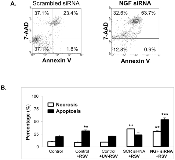
Methodology: Expression of neurotrophic factors (nerve growth factor, NGF; brain-derived neurotrophic factor, BDNF) and receptors (trkA, trkB, p75) was analyzed at the protein level by immunofluorescence and flow cytometry and at the mRNA level by real-time PCR. Targeted siRNA was utilized to blunt NGF expression, and its effect on virus-induced apoptosis/necrosis was evaluated by flow cytometry following annexin V/7-AAD staining.

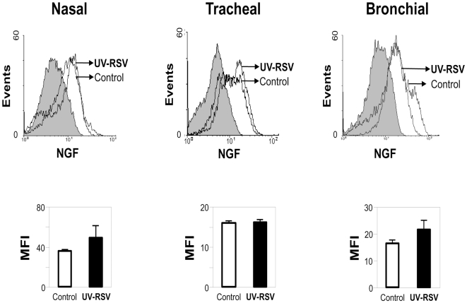
Principal findings: RSV infection was more efficient in cells from more distal (bronchial) vs. more proximal origin. In bronchial cells, RSV infection induced transcript and protein overexpression of NGF and its high-affinity receptor trkA, with concomitant downregulation of the low-affinity p75(NTR). In contrast, tracheal cells exhibited an increase in BDNF, trkA and trkB, and nasal cells increased only trkA. RSV-infected bronchial cells transfected with NGF-specific siRNA exhibited decreased trkA and increased p75(NTR) expression. Furthermore, the survival of bronchial epithelial cells was dramatically decreased when their endogenous NGF supply was depleted prior to RSV infection.

Conclusions/significance: RSV infection of the distal airway epithelium, but not of the more proximal sections, results in overexpression of NGF and its trkA receptor, while the other p75(NTR) receptor is markedly downregulated. This pattern of neurotrophin expression confers protection against virus-induced apoptosis, and its inhibition amplifies programmed cell death in the infected bronchial epithelium. Thus, pharmacologic modulation of NGF expression may offer a promising new approach for management of common respiratory infections.


Conflict of interest statement

Competing Interests: The authors have declared that no competing interests exist.

Figures

Figure 1
Figure 1. RSV infects bronchial epithelial cells more efficiently.
(A) Human nasal, tracheal, and bronchial epithelial cells after infection with GFP-expressing RSV (rgRSV) at 1 MOI for 48 h. The bright field panels (left) show the total number of cells. The green fluorescent cells (center) represent those which are actively infected with RSV. (B) Bronchial epithelial cells are the most susceptible to RSV infection. Flow cytometric data show the percentage of fluorescent (infected) cells in each panel compared to the non-infected control cells (shaded histogram). Data are expressed as the mean±SEM (n = 4 experiments). *** = p<0.001 compared to nasal or tracheal cells.
Figure 2
Figure 2. RSV infection modulates NGF expression.
(A) RSV-induced NGF overexpression in human bronchial epithelial cells infected for 48 h. Cells were incubated with rabbit polyclonal NGF antibody for 1 h and subsequently treated with a secondary PE-conjugated anti-rabbit antibody. Nuclei were stained with TOPRO-3. Pictures were taken using a confocal microscope having a fluorescence emission of 488 nm for detection of green fluorescent protein (GFP). (B) Changes in NGF protein expression were detected by staining rgRSV-infected or non-infected nasal, tracheal and bronchial epithelial cells with anti-human NGF antibody or a matched isotype control (shaded histogram). Primary antibody binding was detected by incubation with PE-conjugated goat anti-rabbit antibody. (C) Geometric mean fluorescent intensity (MFI) is presented in bar graphs. Data are expressed as the mean±SEM (n = 4 experiments). ** = p<0.01 compared to non-infected controls.
Figure 3
Figure 3. UV-inactivated RSV does not change NGF expression.
(A) Following inactivation by UV light, rgRSV lost its effect on NGF expression in human airway epithelial cells. Changes in NGF protein expression were detected by staining UV-RSV-exposed or non-infected nasal, tracheal and bronchial epithelial cells with anti-human NGF antibody or a matched isotype control (shaded histogram). Primary antibody binding was detected by incubation with PE-conjugated goat anti-rabbit antibody. (B) Geometric mean fluorescent intensity (MFI) is presented in bar graphs. Data are expressed as the mean±SEM (n = 4 experiments). None of the effects was statistically significant.
Figure 4
Figure 4. RSV-induced modulation of neurotrophic growth factors and their receptors in (A) nasal epithelial cells; (B) tracheal epithelial cells; (C) bronchial epithelial cells.
Confluent plates of epithelial cells were infected with 1 MOI of rgRSV; after 6 h, the excess viral particles were removed and fresh culture medium added. Subsequently, cells were harvested at 48 h and stained with specific antibody or its matched isotype control (shaded histogram). Cells not exposed to rgRSV were used as controls. Changes in protein expression were measured by flow cytometry using suitable secondary antibodies conjugated with phycoerythrin (PE) or allophycocyanin (APC). Geometric mean fluorescent intensity (MFI) is presented in bar graphs. Data are expressed as the mean±SEM (n = 4 experiments). * = p<0.05; ** = p<0.01; *** = p<0.001 compared to non-infected controls.
Figure 5
Figure 5. RSV-induced changes in neurotrophins gene expression in (A) nasal epithelial cells; (B) tracheal epithelial cells; and (C) bronchial epithelial cells.
Cells were infected with 1 MOI of rgRSV for 24 h; then, RNA was isolated and cDNA synthesized for real-time PCR analysis. Ct values were normalized to the housekeeping gene HPRT1. Data shown are the average of 4 independent experiments performed in duplicate, and are presented as the mean±SEM (n = 8). ** = p<0.01 compared to non-infected controls.
Figure 6
Figure 6. NGF knock-down alters gene expression of other neurotrophins in (A) nasal epithelial cells; (B) tracheal epithelial cells; and (C) bronchial epithelial cells.
After 48 h transfection with either scrambled siRNA (SCRsiRNA) or NGF-specific siRNA (NGFsiRNA), cells were infected with 1 MOI of rgRSV for 24 h. Each experiment included non-transfected and non-infected controls. Transcript levels were analyzed by real-time PCR for relative changes in NGF, BDNF, trkA, trkB, and p75NTR gene expression, and fold change was calculated by normalizing Ct values to HPRT1. Data shown are the average of 3 independent experiments performed in duplicate, and are presented as the mean±SEM (n = 6). ** = p<0.01; *** = p<0.001 compared to matched non-transfected controls.
Figure 7
Figure 7. Apoptosis in RSV-infected bronchial epithelial cells increases after NGF knock-down.
(A) Scrambled siRNA-transfected bronchial cells infected with 1 MOI or 5 MOI of rgRSV. (B) NGF siRNA-transfected bronchial cells infected with 1 MOI or 5 MOI of rgRSV. After 24 h of infection, cells were analyzed for apoptosis by flow cytometry with annexin V PE-conjugated antibody. The shaded portions of the histograms represent matched isotype controls. (C) Geometric mean fluorescence intensity (MFI) in NGF-depleted cells infected with 5 MOI of rgRSV was compared with the scrambled siRNA control. Values were subtracted from the isotype MFI and presented as mean±SEM (n = 4 experiments). ** = p<0.01 compared to controls transfected with scrambled siRNA.
Figure 8
Figure 8. Apoptosis vs. necrosis in RSV-infected bronchial epithelial cells after NGF knock-down.
(A). Dot plots of simultaneous annexin V and 7-aminoactinomycin D (7-AAD) staining of rgRSV-infected bronchial epithelial cells transfected with scrambled (SCR) siRNA (left) or with NGF-specific siRNA (right). After 48 h of transfection, cells were infected with 5 MOI of rgRSV. Apoptosis and necrosis were detected by DNA labeling with 7-AAD-PE and annexin V-Cy3 respectively. Data are expressed as percentage (%) of total cells. (B). Apoptosis vs. necrosis assessed by annexin V and 7-AAD staining followed by flow cytometry. Non-transfected controls include non-infected, rgRSV-infected, and UV-RSV-exposed cells. Data are expressed as the mean±SEM (n = 3 experiments). ** = p<0.01; *** = p<0.001 compared to non-infected controls.
Figure 9
Figure 9. NGF transcripts induction in bronchial epithelial cells exposed to RSV.
(A) RSV transcript levels. (B) NGF transcript levels. Bronchial epithelial cells were infected with 1 MOI of rgRSV. Cells were trypsinized at hourly intervals and cell pellets were collected for RNA extraction and real-time PCR analysis. Data shown are the average of 3 independent experiments performed in duplicate, and are presented as the mean±SEM (n = 6). * = p<0.05; ** = p<0.01; *** = p<0.001 compared to previous time point.

References

    1. Panitch HB. Bronchiolitis in infants. Curr Opin Pediatr. 2001;13:256–260. - PubMed
    1. Piedimonte G. Contribution of neuroimmune mechanisms to airway inflammation and remodeling during and after respiratory syncytial virus infection. Pediatr Infect Dis J. 2003;22:S66–74; discussion S74–65. - PubMed
    1. Psarras S, Papadopoulos NG, Johnston SL. Pathogenesis of respiratory syncytial virus bronchiolitis-related wheezing. Paediatr Respir Rev. 2004;5(Suppl A):S179–184. - PubMed
    1. Steiner RW. Treating acute bronchiolitis associated with RSV. Am Fam Physician. 2004;69:325–330. - PubMed
    1. Openshaw PJ, Dean GS, Culley FJ. Links between respiratory syncytial virus bronchiolitis and childhood asthma: clinical and research approaches. Pediatr Infect Dis J. 2003;22:S58–64; discussion S64–55. - PubMed

Publication types