

Possible brucellosis in an early hominin skeleton from sterkfontein, South Africa
- PMID: 19649274
- PMCID: PMC2713413
- DOI: 10.1371/journal.pone.0006439
Possible brucellosis in an early hominin skeleton from sterkfontein, South Africa
Abstract
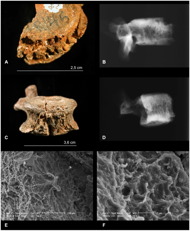
We report on the paleopathological analysis of the partial skeleton of the late Pliocene hominin species Australopithecus africanus Stw 431 from Sterkfontein, South Africa. A previous study noted the presence of lesions on vertebral bodies diagnosed as spondylosis deformans due to trauma. Instead, we suggest that these lesions are pathological changes due to the initial phases of an infectious disease, brucellosis. The macroscopic, microscopic and radiological appearance of the lytic lesions of the lumbar vertebrae is consistent with brucellosis. The hypothesis of brucellosis (most often associated with the consumption of animal proteins) in a 2.4 to 2.8 million year old hominid has a host of important implications for human evolution. The consumption of meat has been regarded an important factor in supporting, directing or altering human evolution. Perhaps the earliest (up to 2.5 million years ago) paleontological evidence for meat eating consists of cut marks on animal remains and stone tools that could have made these marks. Now with the hypothesis of brucellosis in A. africanus, we may have evidence of occasional meat eating directly linked to a fossil hominin.
Conflict of interest statement
Figures

References
-
- Tobias PV. Aspects of pathology and death among early hominids. The Leech. 1974;44:119–124.
-
- Trinkaus E, Zimmerman MR. Trauma among the Shanidar Neandertals. Am J Phys Anthropol. 1982;57:61–76. - PubMed
-
- Cook DC, Buikstra JE, DeRousseau CJ, Johanson DC. Vertebral pathology in the Afar australopithecines. Am J Phys Anthropol. 1983;60:83–101. - PubMed
-
- Lovell NC. An evolutionary framework for assessing illness and injury in nonhuman primates. Am J Phys Anthropol. 1991;34:117–155.
-
- Fisk GR, Macho GA. Evidence of a healed compression fracture in a Plio-Pleistocene hominid talus from Sterkfontein, South Africa. Int J Osteoarch. 1992;2:325–332.
MeSH terms
LinkOut - more resources
Full Text Sources
