. 2010 Jan;119(1):111-22.
doi: 10.1007/s00401-009-0576-2. Epub 2009 Aug 2.

Brain progranulin expression in GRN-associated frontotemporal lobar degeneration

Affiliations

Brain progranulin expression in GRN-associated frontotemporal lobar degeneration

Alice S Chen-Plotkin et al. Acta Neuropathol. 2010 Jan.

Abstract

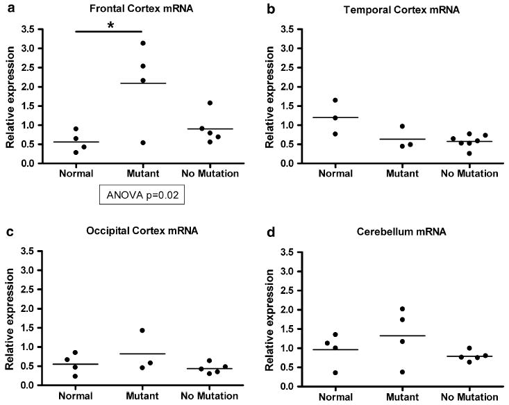
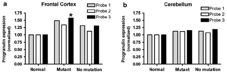
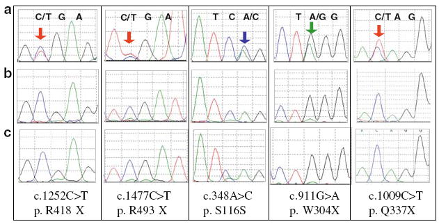
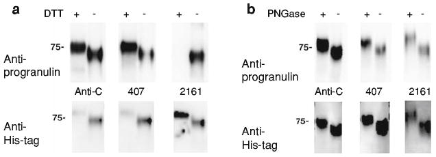
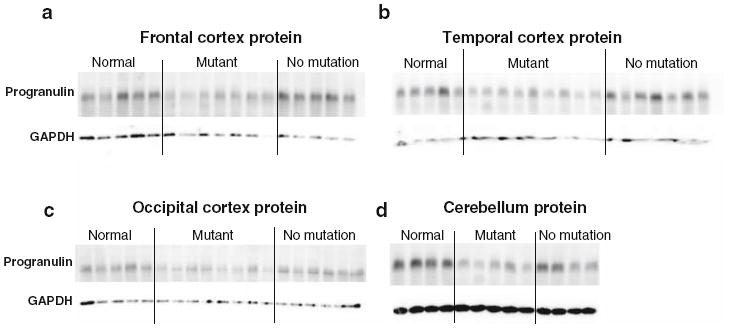
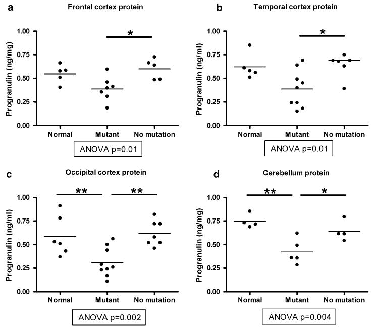
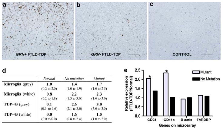
Frontotemporal lobar degeneration with TDP-43 inclusions (FTLD-TDP) is characterized by progressive decline in behavior, executive function, and language. Progranulin (GRN) gene mutations are pathogenic for FTLD-TDP, and GRN transcript haploinsufficiency is the proposed disease mechanism. However, the evidence for this hypothesis comes mainly from blood-derived cells; we measured progranulin expression in brain. We characterized mRNA and protein levels of progranulin from four brain regions (frontal cortex, temporal cortex, occipital cortex, and cerebellum) in FTLD-TDP patients with and without GRN mutations, as well as neurologically normal individuals. Moreover, we performed immunohistochemistry to evaluate the degree of TDP-43 pathology and microglial infiltration present in these groups. In most brain regions, patients with GRN mutations showed mRNA levels comparable to normal controls and to FTLD-TDP without GRN mutations. However, GRN transcript levels in a brain region severely affected by disease (frontal cortex) were increased in mutation-bearing patients. When compared with normal individuals, GRN mutation-bearing cases had a significant reduction in the amount of progranulin protein in the cerebellum and occipital cortex, but not in the frontal and temporal cortices. In GRN mutant cases, GRN mRNA originated from the normal allele, and moderate microglial infiltration was observed. In conclusion, GRN mutation carriers have increased levels of mRNA transcript from the normal allele in brain, and proliferation of microglia likely increases progranulin levels in affected regions of the FTLD-TDP brain, and whether or not these findings underlie the accumulation of TDP-43 pathology in FTLD-TDP linked to GRN mutations remains to be determined.


Conflict of interest statement

Conflict of interest statement The authors declare that they have no conflicts of interest.

Figures

Fig. 1
Fig. 1
Quantification of GRN mRNA transcript levels from brain samples in normal controls (normal), FTLD-TDP with GRN mutations (mutant), and FTLD-TDP without GRN mutations (no mutation). a Quantitative reverse transcription PCR (QRT-PCR) shows that GRN mRNA is increased in frontal cortex samples from FTLD-TDP cases with GRN mutations; increase relative to normal control is significant (P < 0.05). b–d QRT-PCR shows equivalent amounts of temporal cortex, occipital cortex, and cerebellar GRN mRNA in normal controls and FTLD-TDP with and without GRN mutations. QRT-PCR reactions were performed in duplicate, standardized to the geometric mean of two housekeeping genes (cyclophilin A and β-actin), and normalized to control. Individual cases are shown as circles, and the mean for each group is shown as a bar. ANOVA was used to evaluate progranulin mRNA levels across groups, followed by Tukey’s test for pairwise comparisons
Fig. 2
Fig. 2
Microarray analysis corroborates QRT-PCR results.a Microarray analysis demonstrates that frontal cortex from GRN mutants (mutant, n = 6) have elevated GRN mRNA relative to controls (normal, n = 8) and FTLD-TDP without GRN mutations (no mutation, n = 10). GRN is represented by three different probes on the Affymetrix U133A microarray used. For probe 3 (*), GRN mutation cases show a significant elevation (P = 0.04) relative to normals. P value corrected for multiple-hypothesis testing by the Benjamini–Hochberg method. b Cerebellar samples from normal controls (n = 8), FTLD-TDP with GRN mutations (n = 5), and FTLD-TDP without GRN mutations (n = 8) show equivalent amounts of GRN mRNA by microarray
Fig. 3
Fig. 3
Sequencing of cDNA reveals that increased mRNA transcript levels originate from the normal allele. Brain mRNA from GRN mutants was reverse-transcribed, and the resulting cDNA was sequenced. Row A depicts genomic DNA for each GRN mutant; mutant peaks are indicated by colored arrows. Row B depicts cDNA for each GRN mutant, and Row C depicts normal cDNA for comparison. For all GRN mutants tested, mRNA primarily represents the normal allele indicating that the increased transcript levels result from the normal allele
Fig. 4
Fig. 4
Novel antibodies recognize reduced and non-reduced forms of progranulin. We generated antibodies recognizing different forms of human progranulin. a Purified His-tagged human progranulin treated (+) or untreated (−) with DTT, a reducing agent, was separated on a 7.5% SDS-PAGE gel, and blotted with antibodies indicated (top). The blot was then stripped and re-blotted with anti-His antibody (bottom). As shown in the top, the rabbit polyclonal anti-progranulin Ct antibody (anti-C) does not discriminate between native (no DTT treatment) and reduced (DTT treatment) forms of progranulin, while mAb 407 (407) preferentially recognizes reduced progranulin, and mAb 2161 (2161) recognizes native progranulin. The native non-reduced form of progranulin runs faster relative to the reduced form of progranulin due to the compact conformation of the native protein. b Purified His-tagged human progranulin was deglycosylated with PNGase F (+) or mock-treated (−), and proteins were separated on a 7.5% SDS-PAGE gel. Immunoblotting with rabbit anti-C, mAb 407, or mAb 2161 demonstrates that in each case deglycosylation results in a decrease in the apparent molecular weight of progranulin, consistent with the nature of progranulin as a highly glycosylated protein. As before, immunoblots were performed with the labeled antibody (top), followed by stripping and re-blotting with anti-His antibody (bottom). Because of preferential binding for reduced or non-reduced progranulin, DTT-treated progranulin was used for rabbit anti-C and mAb 407 immunoblots in (b), while non-treated progranulin was used for mAb 2161 immunoblotting
Fig. 5
Fig. 5
Immunoblot of progranulin protein from brain samples in normal controls (normal), FTLD-TDP with GRN mutations (mutant), and FTLD-TDP without GRN mutations (no mutation). Representative immunoblot of progranulin protein from frontal cortex (a), temporal cortex (b), occipital cortex (c), and cerebellum (d) using mAb 407. In brain regions relatively unaffected by disease (occipital cortex and cerebellum), GRN mutants show uniformly decreased progranulin levels relative to neurologically normal controls, while brain regions that are histopathologically affected in FTLD-TDP (frontal cortex and temporal cortex) show more individual to individual variation in progranulin protein levels among GRN mutants. Each lane represents a separate patient or control sample. Immunoblots were repeated four times. Human brain lysates (RIPA fraction) were separated on a 7.5% SDS-PAGE gel
Fig. 6
Fig. 6
Sandwich ELISA quantification of progranulin protein levels from brain samples in normal controls (normal), FTLD-TDP with GRN mutations (mutant), and FTLD-TDP without GRN mutations (no mutation). Progranulin in human brain lysates (RIPA fraction) was captured with mAb 2161 and detected with rabbit anti-progranulin Ct antibody and HRP-conjugated goat anti-rabbit antibody. a, b In brain regions with more histopathological involvement in FTLD-TDP (frontal and temporal cortex), ELISA shows no significant difference in progranulin protein levels between normal controls and GRN mutants, although GRN mutants did differ from FTLD-TDP without GRN mutations (see bars with * comparing FTLD-TDP cases ± GRN mutations). c, d In brain regions that are relatively histopathologically spared in FTLD-TDP (occipital cortex and cerebellum), ELISA shows significantly decreased progranulin protein levels in GRN mutants relative to both normal controls and FTLD-TDP without GRN mutations (see bars with * or ** comparing GRN mutants to normal controls or FTLD-TDP cases without GRN mutations). Individual cases are shown as circles, and the mean for each group is shown as a bar. ANOVA was used to evaluate progranulin protein levels across groups, followed by Tukey’s test for pairwise comparisons *P < 0.05, **P < 0.01
Fig. 7
Fig. 7
Microglial infiltration in GRN mutants. a–c Gray matter sections from frontal cortex are shown for cases of (left to right) FTLD-TDP with GRN mutations (GRN+), FTLD-TDP without GRN mutations (GRN–), and normal controls. Cases stained for microglia with HLA-DR. Scale bar represents 200 μm. d Scoring of immunohistochemical staining for TDP-43 pathology and microglial infiltration was performed for frontal cortex sections by a neuropathologist blinded to disease and GRN mutation status. Scores ranged from 0 (no microglial staining) to 3 (most robust microglial staining). GRN+ FTLD-TDP showed more microglial infiltration than GRN– FTLD- TDP and normals in both gray and white matter. Values shown are median score and interquartile range (parentheses) calculated from grouped data. e Microarray expression results are shown for two microglial markers (CD34 and CD11b) as well as a housekeeping gene (β-actin) and the gene encoding TDP-43 (TARDBP). Significantly increased mRNA expression was seen in frontal cortex samples from GRN mutants for CD34 (*P = 0.02) and CD11b (*P = 0.05). Bars represent relative expression for FTLD-TDP with (GRN mutant) and without (no mutation) GRN mutations, normalized to control. P values are corrected for multiple-hypothesis testing

Comment in

References

    1. Akiyama H, McGeer PL. Brain microglia constitutively express beta-2 integrins. J Neuroimmunol. 1990;30:81–93. - PubMed
    1. Arai T, Hasegawa M, Akiyama H, et al. TDP-43 is a component of ubiquitin-positive tau-negative inclusions in frontotemporal lobar degeneration and amyotrophic lateral sclerosis. Biochem Biophys Res Commun. 2006;351:602–611. - PubMed
    1. Asheuer M, Pflumio F, Benhamida S, et al. Human CD34+ cells differentiate into microglia and express recombinant therapeutic protein. Proc Natl Acad Sci USA. 2004;101:3557–3562. - PMC - PubMed
    1. Baker CA, Manuelidis L. Unique inflammatory RNA profiles of microglia in Creutzfeldt–Jakob disease. Proc Natl Acad Sci USA. 2003;100:675–679. - PMC - PubMed
    1. Baker M, Mackenzie IR, Pickering-Brown SM, et al. Mutations in progranulin cause tau-negative frontotemporal dementia linked to chromosome 17. Nature. 2006;442:916–919. - PubMed

Publication types

MeSH terms