. 2009 Oct;27(10):2614-23.
doi: 10.1002/stem.187.

Direct evidence of mesenchymal stem cell tropism for tumor and wounding microenvironments using in vivo bioluminescent imaging

Affiliations

Direct evidence of mesenchymal stem cell tropism for tumor and wounding microenvironments using in vivo bioluminescent imaging

Shannon Kidd et al. Stem Cells. 2009 Oct.

Abstract

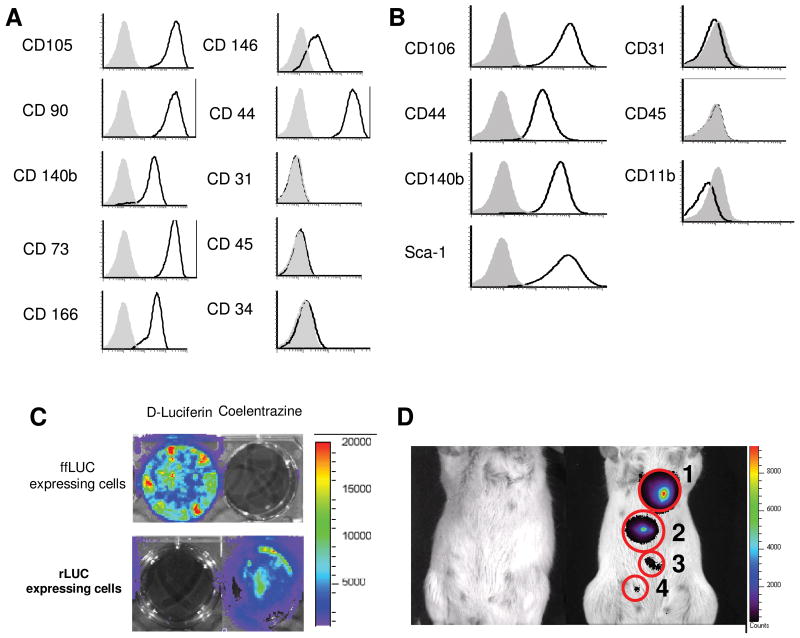
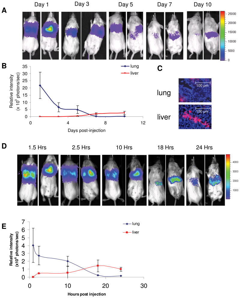
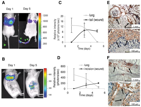
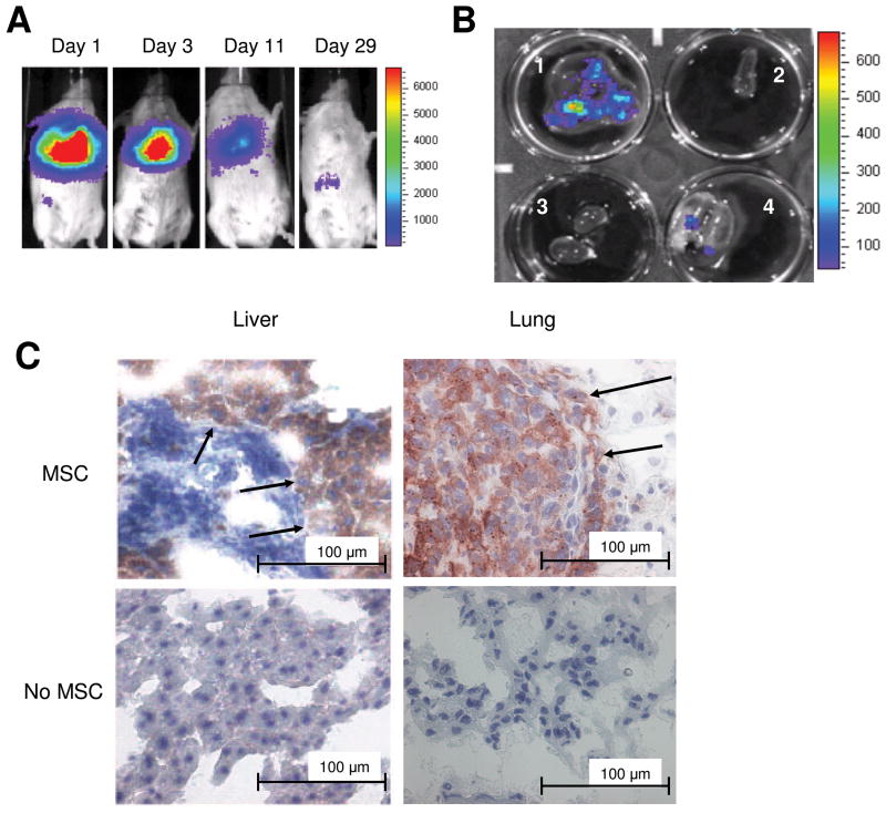
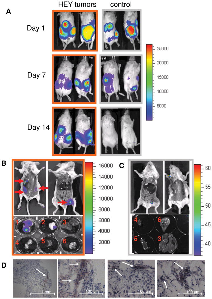
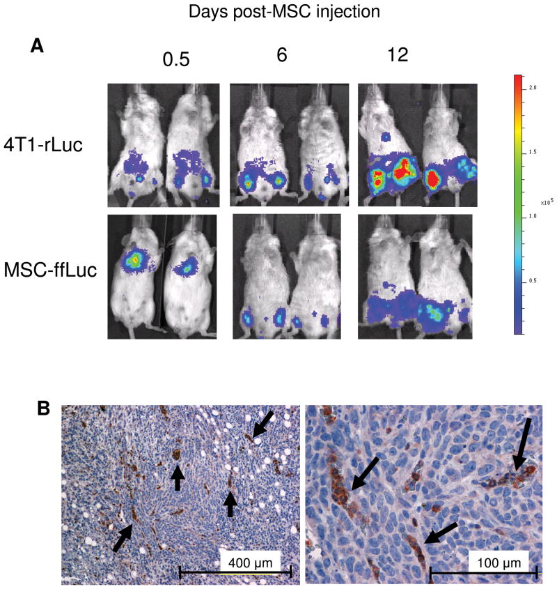
Multipotent mesenchymal stromal/stem cells (MSC) have shown potential clinical utility. However, previous assessments of MSC behavior in recipients have relied on visual detection in host tissue following sacrifice, failing to monitor in vivo MSC dispersion in a single animal and limiting the number of variables that can be observed concurrently. In this study, we used noninvasive, in vivo bioluminescent imaging to determine conditions under which MSC selectively engraft in sites of inflammation. MSC modified to express firefly luciferase (ffLuc-MSC) were injected into healthy mice or mice bearing inflammatory insults, and MSC localization was followed with bioluminescent imaging. The inflammatory insults investigated included cutaneous needle-stick and surgical incision wounds, as well as xenogeneic and syngeneic tumors. We also compared tumor models in which MSC were i.v. or i.p. delivered. Our results demonstrate that ffLuc-expressing human MSC (hMSC) systemically delivered to nontumor-bearing animals initially reside in the lungs, then egress to the liver and spleen, and decrease in signal over time. However, hMSC in wounded mice engraft and remain detectable only at injured sites. Similarly, in syngeneic and xenogeneic breast carcinoma-bearing mice, bioluminescent detection of systemically delivered MSC revealed persistent, specific colocalization with sites of tumor development. This pattern of tropism was also observed in an ovarian tumor model in which MSC were i.p. injected. In this study, we identified conditions under which MSC tropism and selective engraftment in sites of inflammation can be monitored by bioluminescent imaging over time. Importantly, these consistent findings were independent of tumor type, immunocompetence, and route of MSC delivery.


Figures

Figure 1
Figure 1
MSC characterization and labeling for in vivo visualization. (A) hMSC and (B) mMSC were evaluated for phenotypic markers by flow cytometry. (C)ffLuc-expressing and rLuc-expressing cells showed specific reactivity with D-Luciferin and coelentrazine, respectively. (D) In vivo detection of MSC-ffLuc was evaluated at 0 (far left mouse), (1) 300, (2) 100, (3) 30, and (4) 10 cells per sub cutaneous injection. Increasing numbers of MSC demonstrated correlating increases in signal intensity.
Figure 2
Figure 2
Biodistribution of MSC in homeostatic animals. hMSC were IV injected into SCID mice (n=5) and (A) imaged days 1, 3, 5, 7 and 10 post-injection, showing initial localization in the lung and eventual localization in the liver and spleen by Day 10. (B) Bioluminescent signal was quantified in the lung and liver/spleen regions. (C) DiI labeled hMSC were detected in the liver and lung on Day 7 (20x magnification). In a syngeneic model, Balb/C mMSC-ffLuc were IV injected into homeostatic Balb/C mice (n=5) and (D) imaged 1.5, 2.5, 10, 18 and 24 hours post-injection. (E) The bioluminescent signal was quantified over this time indicated initial mMSC distribution in the lung that had fully migrated to the liver/spleen by 24 hours post injection.
Figure 3
Figure 3
hMSC co-localization with subcutaneous wound models. hMSC-ffLuc were injected immediately post needle puncture (Day 1) or 3 days post-surgical incision (Day 1). In the needle puncture model, hMSC-ffLuc were injected into SCID mice immediately post-wound infliction. Images are shown for representative animals at 1 and 5 days (A) post-needle puncture (n=5) and (B) post-lateral incision (n=3). Bioluminescent activity was quantified on days 1, 3, and 5, demonstrating a decrease in activity in the lung and concurrent increases of activity in the (C) tail wounds and (D) cutaneous incisions. IHC on sections of (E) tail and (F) wounded skin demonstrated incorporation of ff-Luc+ hMSC.
Figure 4
Figure 4
hMSC co-localization with MDA-231 breast cancer in SCID mice. MDA-MB-231 cells were IV injected into SCID mice (n=5).
Figure 5
Figure 5
MSC tropism for HEY ovarian carcinoma. SCID mice were IP injected with HEY cells (n=3; orange outline) or PBS (n=3; grey outline). 15 days later, hMSC-ffLuc were IP injected in tumor-bearing and control mice (Day 1). (A) Images were acquired at days 1, 7, and 14 indicating initial dissemination throughout the peritoneal cavity, followed by specific localization in tumor-bearing animals and disappearance in control animals. On day 14, the mice were sacrificed bioluminescent activity was localized to sites of visible tumor development in the open cavities and dissected organs [(1) ventral tumor, (2) dorsal tumor, (3) liver, (4) kidney, (5) spleen, and (6) heart and lungs] of (B) HEY-bearing but not in (C) control mice. (D) IHC for ffLuc on tumor sections from the HEY-bearing mice confirmed the presence of hMSC (magnification as indicated).
Figure 6
Figure 6
mMSC co-localize with subcutaneous, syngeneic 4T1 murine breast carcinoma. 4T1 cells were subcutaneously injected into the hind limbs of Balb/C mice (n=5). 10 days post tumor establishment, mMSC-ffLuc were IV injected. (A) Mice were imaged 0.5, 6 and 12 days post mMSC-ffLuc injection for rLuc (tumor) and ffLuc (mMSC) activities, demonstrating co-localization at days 6 and 12. (B) IHC on 4T1 tumor sections revealed incorporation of ffLuc+MSC.

References

    1. Nakamizo A, Marini F, Amano T, et al. Human bone marrow-derived mesenchymal stem cells in the treatment of gliomas. Cancer Res. 2005;65:3307–3318. - PubMed
    1. Studeny M, Marini FC, Dembinski JL, et al. Mesenchymal stem cells: potential precursors for tumor stroma and targeted-delivery vehicles for anticancer agents. J Natl Cancer Inst. 2004;96:1593–1603. - PubMed
    1. Sasaki M, Abe R, Fujita Y, et al. Mesenchymal stem cells are recruited into wounded skin and contribute to wound repair by transdifferentiation into multiple skin cell type. J Immunol. 2008;180:2581–2587. - PubMed
    1. Satake K, Lou J, Lenke LG. Migration of mesenchymal stem cells through cerebrospinal fluid into injured spinal cord tissue. Spine. 2004;29:1971–1979. - PubMed
    1. Neuhuber B, Timothy Himes B, Shumsky JS, et al. Axon growth and recovery of function supported by human bone marrow stromal cells in the injured spinal cord exhibit donor variations. Brain Res. 2005;1035:73–85. - PubMed

Publication types

MeSH terms