Review
. 2009;46(4):190-209.
doi: 10.1080/10408360902982128.

Vitamin D and differentiation in cancer

Affiliations
Review

Vitamin D and differentiation in cancer

Elzbieta Gocek et al. Crit Rev Clin Lab Sci. 2009.

Abstract

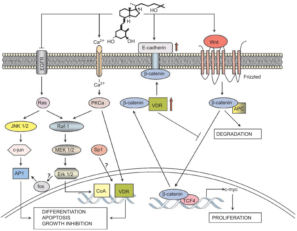
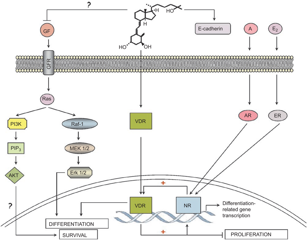
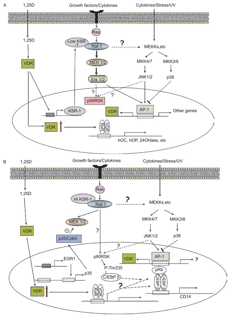
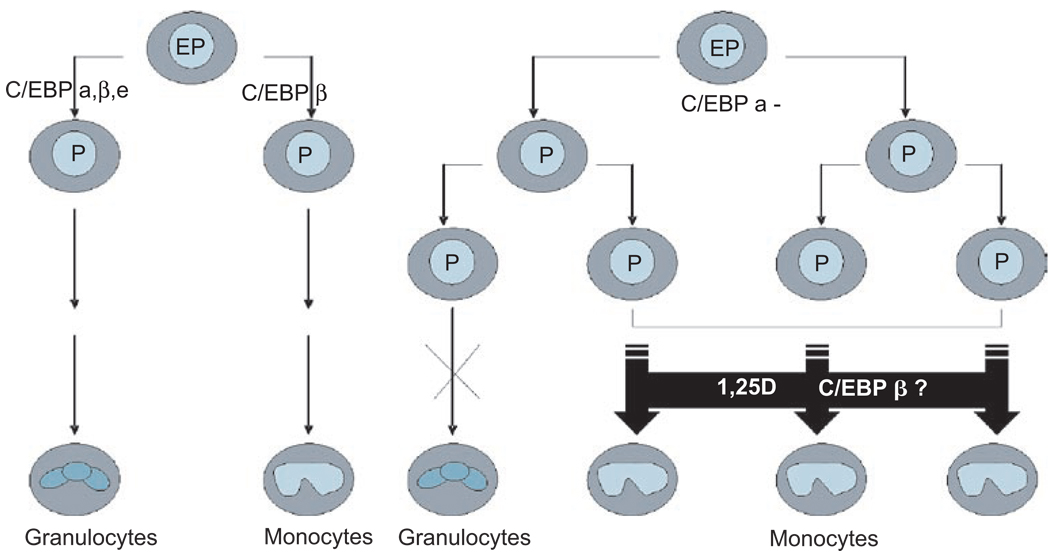
This paper reviews the current understanding of the vitamin D-induced differentiation of neoplastic cells, which results in the generation of cells that acquire near-normal, mature phenotype. Examples of the criteria by which differentiation is recognized in each cell type are provided, and only those effects of 1alpha,25-dihydroxyvitamin D(3) (1,25D) on cell proliferation and survival that are associated with the differentiation process are emphasized. The existing knowledge, often fragmentary, of the signaling pathways that lead to vitamin D-induced differentiation of colon, breast, prostate, squamous cell carcinoma, osteosarcoma, and myeloid leukemia cancer cells is outlined. The important distinctions between the different mechanisms of 1,25D-induced differentiation that are cell-type and cell-context specific are pointed out where known. There is a considerable body of evidence that the principal human cancer cells can be suitable candidates for chemoprevention or differentiation therapy with vitamin D. However, further studies are needed to fully understand the underlying mechanisms in order to improve the therapeutic approaches.


Figures

Figure 1
Figure 1
The suggested pathways of 1,25D-induced differentiation in colon cancer. In proliferating colon epithelial cells the β-catenin complexed with TCF-4 drives the expression of growth promoting genes such as c-myc. This is under the control of Wnt and its surface receptor Frizzled, which inactivate GSK-3β (not shown) and allow the accumulation of β-catenin and thus growth promotion. Binding of β-catenin by VDR, or by other proteins, including E-cadherin, the expression of which is induced by 1,25D (formula shown) leads to the loss of β-catenin from the transcriptional complex in the nucleus, and, as a consequence, to decreased cell proliferation. Also shown is the activation of PKCα by 1,25D-induced influx of calcium (Ca2+), which can activate by phosphorylation the transcriptional activity of VDR and repression of EGFR by 1,25D in colon-derived cells.
Figure 2
Figure 2
Signaling of differentiation by 1,25D in hormone-dependent cancer cells. This schematic illustrates the hypothesis that in normal breast or prostate cells, estrogen (E2) or androgen (A) is sufficient to induce differentiation, respectively. In cancer cells the differentiation signal provided by the hormone-liganded nuclear receptor (NR) may need to be amplified by the cooperation with 1,25D-activated VDR to induce differentiation. Since cells also receive signals from growth factors (GF), several of which activate Ras, the presence of a Ras-activated signaling pathways is exemplified by the AKT and ERK cascades, though the role of these pathways in the differentiation of hormone-dependent cells is uncertain.
Figure 3
Figure 3
(A) Suggested signaling of the early stages of 1,25D-induced monocytic differentiation. Binding of 1,25D to VDR stimulates its translocation to the cell nucleus where it heterodimerizes with RXR, and in myeloid precursor cells, it transactivates genes containing VDREs in their promoter regions. These include genes that encode proteins involved in calcium homeostasis and bone integrity, such as osteocalcin (hOC), osteopontin (hOP), and the 1,25D-catabolic enzyme 24-hydroxylase (24OHase). It is postulated that the regulators of signaling pathways, e.g. KSR-1, are also up-regulated in myeloid cells and alter Ras signaling from the cell membrane so that signaling by MAPKs (MEKs, ERKs, and JNKs) increases the AP-1 activity. This can have a positive feedback effect on differentiation by increasing VDR abundance. It is also suggested that a potential negative feedback mechanism is provided by p38 MAPK, as inhibition of its signaling by SB203580 enhances 1,25D-induced monocytic differentiation. (B) Later stages of 1,25D-induced differentiation. This figure illustrates that the transcription factor EGR-1, known to be up-regulated by 1,25D (189), can increase the expression of p35/Nck5a (p35) activator of Cdk5. Cdk5 activated by p35 then can phosphorylate MEK on Thr286, a site that inactivates it (200), as shown by the Θ symbol. This diminishes ERK1/2 activity downstream from MEK (not shown here), but Raf-1 can activate p90RSK directly, which, in turn, activates the transcription factor C/EBP β, perhaps bound to pRb, and increases the expression of CD14, as part of monocytic differentiation. The activation of p90RSK may also be increased by the JNK pathway, which also activates AP-1, and may lead to VDR expression. The interplay between the signaling by 1,25D, growth factor, and stress add to the overall complexity of the induction of the monocytic phenotype.
Figure 4
Figure 4
The suggested role of CAAT/Enhancer Binding Protein β in 1,25D-induced bypass of the differentiation block in leukemia cells. In this scenario, C/EBP α is indispensable for normal granulopoiesis, while C/EBP β regulates monocytic differentiation. When C/EBP α is mutated or inactivated and granulopoiesis is blocked, immature myeloid cells accumulate in the bone marrow and appear in the peripheral blood, resulting in AML. 1,25D-induced expression of C/EBP β may allow the cells to bypass this block to granulocytic differentiation by switching the lineage of cell differentiation from granulocytes to monocytes.

References

    1. Smithers DW. Maturation in human tumours. Lancet. 1969;2:949–952. - PubMed
    1. Walton JD, Kattan DR, Thomas SK, Spengler BA, Guo HF, Biedler JL, Cheung NK, Ross RA. Characteristics of stem cells from human neuroblastoma cell lines and in tumors. Neoplasia. 2004;6:838–845. - PMC - PubMed
    1. Chen SJ, Zhu YJ, Tong JH, Dong S, Huang W, Chen Y, Xiang WM, Zhang L, Li XS, Qian GQ. Rearrangements in the second intron of the RARalfa gene are present in a large majority of patients with acute promyelocytic leukemia and are used as molecular marker for retinoic acid-induced leukemic cell differentiation. Blood. 1991;78:2696–2701. - PubMed
    1. Degos L, Wang ZY. All trans retinoic acid in acute promyelocytic leukemia. Oncogene. 2001;20:7140–7145. - PubMed
    1. Schlenk RF, Frohling S, Hartmann F, Fischer JT, Glasmacher A, del Valle F, Grimminger W, Gotze K, Waterhouse C, Schoch R, Pralle H, Mergenthaler HG, Hensel M, Koller E, Kirchen H, Preiss J, Salwender H, Biedermann HG, Kremers S, Griesinger F, Benner A, Addamo B, Dohner K, Haas R, Dohner H. Phase III study of all-trans retinoic acid in previously untreated patients 61 years or older with acute myeloid leukemia. Leukemia. 2004;18:1798–1803. - PubMed

Publication types

LinkOut - more resources