. 2010 Oct;14(10):2470-82.
doi: 10.1111/j.1582-4934.2009.00863.x.

Thymic involution, a co-morbidity factor in amyotrophic lateral sclerosis

Affiliations

Thymic involution, a co-morbidity factor in amyotrophic lateral sclerosis

Akop Seksenyan et al. J Cell Mol Med. 2010 Oct.

Abstract

Amyotrophic lateral sclerosis (ALS) is a devastating disease, characterized by extremely rapid loss of motor neurons. Our studies over the last decade have established CD4(+) T cells as important players in central nervous system maintenance and repair. Those results, together with recent findings that CD4(+) T cells play a protective role in mouse models of ALS, led us to the current hypothesis that in ALS, a rapid T-cell malfunction may develop in parallel to the motor neuron dysfunction. Here, we tested this hypothesis by assessing thymic function, which serves as a measure of peripheral T-cell availability, in an animal model of ALS (mSOD1 [superoxide dismutase] mice; G93A) and in human patients. We found a significant reduction in thymic progenitor-cell content, and abnormal thymic histology in 3-4-month-old mSOD1 mice. In ALS patients, we found a decline in thymic output, manifested in the reduction in blood levels of T-cell receptor rearrangement excision circles, a non-invasive measure of thymic function, and demonstrated a restricted T-cell repertoire. The morbidity of the peripheral immune cells was also manifested in the increase of pro-apoptotic BAX/BCXL2 expression ratio in peripheral blood mononuclear cells (PBMCs) of these patients. In addition, gene expression screening in the same PBMCs, revealed in the ALS patients a reduction in key genes known to be associated with T-cell activity, including: CD80, CD86, IFNG and IL18. In light of the reported beneficial role of T cells in animal models of ALS, the present observation of thymic dysfunction, both in human patients and in an animal model, might be a co-pathological factor in ALS, regardless of the disease aetiology. These findings may lead to the development of novel therapeutic approaches directed at overcoming the thymic defect and T-cell deficiency.


Figures

Fig 1
Fig 1
Thymic cell content is reduced in mSOD-1 mice during the course of disease progression. Two-colour flow-cytometry was used to analyse the phenotypes and total cell number in the thymus of mSOD1 mice and their age-matched wild-type controls during the progressive stage of the disease (day 120; A–G) and prior to disease onset (day 60; H). (A–E). Total thymocyte number is significantly reduced at the progressive stage of the disease: (A) Total number of thymocytes (t12=−2.84; *P= 0.01). (B) Total number of DN thymocytes (t12=−2.12; *P= 0.05). (C) Total number of DP thymocytes (t12=−2.97; P=*0.01). (D) Total number of CD4+ cells (t12=−2.07; P= 0.06). (E) Total number of CD8+ cells (t12=−2.2; P=*0.04). Reduction in thymic cellularity correlated with an increased proportion of DN cells (F, t6.4= 2.82; *P= 0.02) and decreased proportion of DP cells (G, t6.6=−2.5; *P= 0.04). (H) Significant elevation in the absolute number of CD8 cells was observed at day 60, prior to disease onset (t4.23= 2.7; *P= 0.05). Values represent means ± S.E.M. (I) Representative photomicrographs of thymus collected from mSOD-1 mouse at the end-stage of the disease and its age-matched wild-type control. (J) Thymic dysplasia was detected in mSOD1 mice upon disease progression. Sections taken through the thymus were stained by haematoxylin and eosin. (Ji) A representative thymic section derived from a 60-day-old wild-type mouse. Note the distinct separation between the cortex (C) and the medulla (M). (Jii) Micrograph showing a higher magnification of the indicated area. (Jiii) A representative thymic section derived from 60-day-old mSOD1 mouse, before disease onset. A normal structure is still observed at this time-point. (Jiv) A representative thymic section derived from 120-day-old mSOD1 mouse. No clearly delineated cortex and medulla can be discerned at this stage. Bars represent 100 μm. (K) Vβ T-cell repertoire in the peripheral T-cell pool of mSOD1 and control mice. No major changes in T-cell clonality were observed in mSOD-1 mice compared to age-matched wild-type mice, along disease progression.
Fig 2
Fig 2
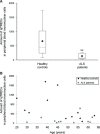
Thymic output is severely reduced in ALS patients. Total DNA was purified from PBMCs that were collected from 11 ALS patients (30–62 years old; average age: 49.1 ± 10.3) and 12 age-matched (25–63 years old; average age: 45.1 ± 12.3; P= 0.40) healthy controls. (A) The amount of sjTREC was measured by Q-PCR, and was normalized against RNAseP. A significant reduction was found in the copies of sjTREC in the blood of ALS patients, compared to controls (t13.4=−3.06; **P= 0.008). The graph shows the interquartile range of the measurements. The dashed line represents the median value; the circle indicates the average value. (B) Scatterplot of sjTREC levels by age.
Fig 3
Fig 3
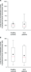
Normal levels of early T-cell recombination events in ALS patients. Total DNA was purified from PBMCs that were collected from 11 ALS patients (30–62 years old; average age: 49.1 ± 10.3) and 12 age-matched (25–63 years old; average age: 45.1 ± 12.3; P= 0.40) control donors. The early recombination events were quantified using Q-PCR. No differences were found in the two tested events, D-D (A; t21=−0.15; P= 0.88) and D-J (B; t21=−0.35; P= 0.73). The graphs indicate the interquartile range of the measurements. The dashed line represents the median value; the circle indicates the average value.
Fig 4
Fig 4
Restricted TCRγ repertoire in ALS patients. Genescan display of four PCR-generated profiles of TCRγ rearrangements, from five representative patients. Vγ9/2 and Vγfl represent FAM labelled fluorescent variable region primers. Vγ11 and Vγ10/2 are HEX labelled fluorescent variable region primers. Nine additional patients were similarly analysed and demonstrated a similar profile, suggesting restricted polyclonality, though not monoclonality, relative to normal healthy individuals.
Fig 5
Fig 5
Reduced immune potential and increased susceptibility to apoptosis in PBMCs from ALS patients. Expression levels of (A) CD80 (Z= 2.3, *P= 0.02), (B) CD86 (Z= 3.0, **P= 0.002), (C) IL8 (Z=−2.5, *P= 0.01), (D) IFNG (Z= 2.8, **P= 0.006), (E) IL18 (Z= 3.0, **P= 0.002) and (F) BAX to BCL2 ratio (Z=−3.0, **P= 0.002). Expression was assessed by real-time Q-PCR. Values represent expression levels in arbitrary units, normalized against 18S in the same samples (n= 8 and n= 6 for ALS patients and healthy controls, respectively; each was tested in triplicate). Error bars represent S.D.

Similar articles

Cited by

References

    1. Lee JR, Annegers JF, Appel SH. Prognosis of amyotrophic lateral sclerosis and the effect of referral selection. J Neurol Sci. 1995;132:207–15. - PubMed
    1. Rosen DR. Mutations in Cu/Zn superoxide dismutase gene are associated with familial amyotrophic lateral sclerosis. Nature. 1993;362:59–62. - PubMed
    1. Boillee S, Yamanaka K, Lobsiger CS, et al. Onset and progression in inherited ALS determined by motor neurons and microglia. Science. 2006;312:1389–92. - PubMed
    1. Clement AM, Nguyen MD, Roberts EA, et al. Wild-type nonneuronal cells extend survival of SOD1 mutant motor neurons in ALS mice. Science. 2003;302:113–7. - PubMed
    1. Gowing G, Philips T, Van Wijmeersch B, et al. Ablation of proliferating microglia does not affect motor neuron degeneration in amyotrophic lateral sclerosis caused by mutant superoxide dismutase. J Neurosci. 2008;28:10234–44. - PMC - PubMed

Publication types